An overview of the immune system, detailing the innate and adaptive responses, B cells, T cells, NK cells, and the roles of MHC class I and II molecules in antigen presentation and immune regulation.
The immune system is a complex, highly coordinated network of cells, tissues, and organs designed to protect the body from pathogenic invasion and internal malfunctions, such as tumor growth. To understand how the body distinguishes "self" from "non-self," we must examine the two primary arms of the immune response: the innate and the adaptive systems. While one provides immediate, non-specific defense, the other offers a highly refined, memory-based response tailored to specific threats.
Innate vs. Adaptive Immunity
The immune response is broadly categorized into two systems that work in tandem.
Innate Immunity
Innate immunity serves as the body's first line of defense. It is characterized by a rapid response—occurring within minutes to hours—and utilizes germline-encoded mechanisms that do not change with repeated exposure.
Components: Neutrophils, macrophages, monocytes, dendritic cells, natural killer (NK) cells, complement proteins, and physical barriers like the skin and epithelial linings.
Recognition: It relies on pattern recognition receptors, such as Toll-like receptors (TLRs), which identify pathogen-associated molecular patterns (PAMPs). Common PAMPs include lipopolysaccharide (LPS) from gram-negative bacteria and viral nucleic acids.
Adaptive Immunity
Adaptive immunity is highly specific and develops over a longer period. Unlike the innate system, it utilizes V(D)J recombination during lymphocyte development to create a vast array of unique receptors.
Components: B cells, T cells, and circulating antibodies.
Memory: A hallmark of adaptive immunity is the creation of memory cells. Upon subsequent exposure to the same antigen, the immune response is significantly faster and more robust.
The Adaptive Response: Humoral and Cell-Mediated Immunity
The adaptive immune system is divided into two functional arms: humoral immunity and cell-mediated immunity.
Humoral Immunity (B Cells)
Humoral immunity is the "antibody-producing arm" of the adaptive response. It is primarily mediated by B cells.
Antigen Recognition: B cells recognize and present antigens.
Optimization: Through a process called somatic hypermutation, B cells optimize their antigen specificity.
Effector Function: Once activated, B cells differentiate into plasma cells, which secrete specific immunoglobulins (antibodies) to neutralize pathogens.
Memory: Memory B cells persist in the body to accelerate future responses.
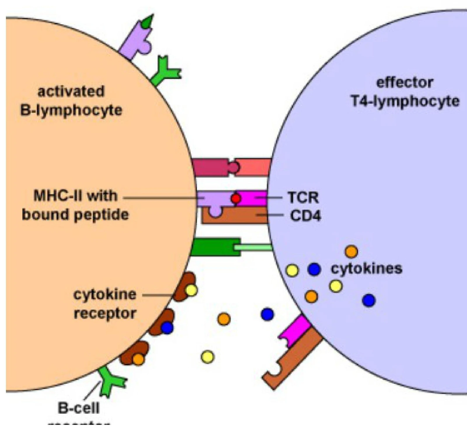
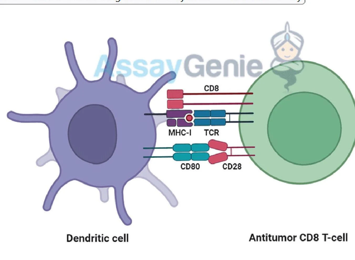
Diagram illustrating the interaction between an activated B-lymphocyte and an effector T4-lymphocyte, showing the roles of MHC-II, TCR, CD4, and cytokines in cell communication.Diagram illustrating the interaction between a dendritic cell and an antitumor CD8 T-cell, showing key molecular components like MHC-I, TCR, CD8, CD80, and CD28.
Cell-Mediated Immunity (T Cells)
Cell-mediated immunity does not involve antibodies but instead relies on T cells to destroy infected or abnormal cells. There are two major subsets of T cells:
CD4+ Helper T Cells: These cells act as the "generals" of the immune system. They help B cells produce antibodies and secrete cytokines to recruit phagocytes and activate other leukocytes.
CD8+ Cytotoxic T Cells: These cells are the "soldiers" that directly kill virus-infected or tumor cells. They induce apoptosis (programmed cell death) using perforin and granzymes.
The Rule of 8
A useful mnemonic to remember the relationship between MHC molecules and T cell subsets is the "Rule of 8":
MHC II×CD4=8
MHC I×CD8=8
Antigen Presentation and MHC Molecules
For T cells to recognize an antigen, the antigen must be "presented" on the surface of a cell via Major Histocompatibility Complex (MHC) molecules. In humans, MHC genes are known as HLA (Human Leukocyte Antigen) genes.
MHC Class I
MHC I molecules are expressed on all nucleated cells (except red blood cells).
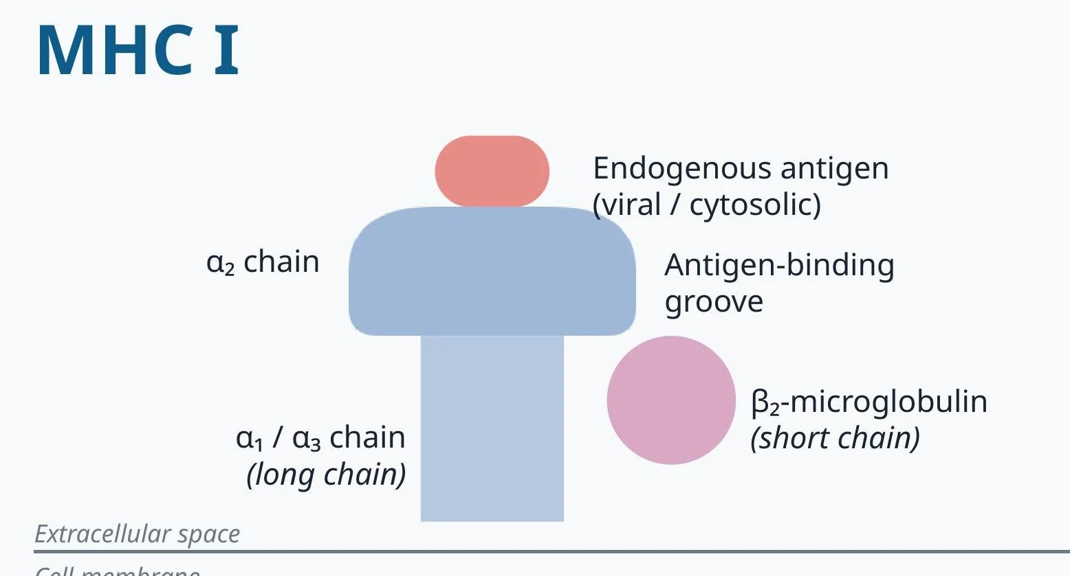
Function: They present endogenous antigens (proteins produced inside the cell, such as viral or tumor proteins) to CD8+ cytotoxic T cells.
Structure: Consists of one long α chain and one short β2-microglobulin chain.
Loading: Antigens are loaded in the Rough Endoplasmic Reticulum (RER) via the TAP transporter.
Diagram illustrating the structure of MHC Class I, showing its alpha chains, beta-2 microglobulin, and the antigen-binding groove which presents endogenous antigens.
MHC Class II
MHC II molecules are expressed primarily on Antigen-Presenting Cells (APCs), such as dendritic cells, macrophages, and B cells.
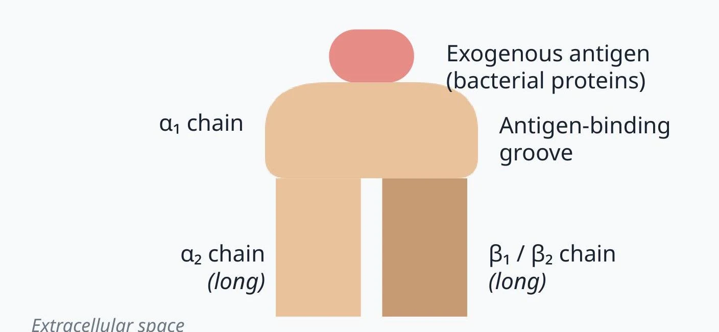
Function: They present exogenous antigens (proteins taken up from outside the cell, such as bacteria) to CD4+ helper T cells.
Structure: Consists of two equal-length chains (α and β).
Loading: Antigens are loaded in an acidified endosome following the release of an invariant chain.
Diagram illustrating the structure of a major histocompatibility complex (MHC) class I molecule, showing its alpha chains and beta-2 microglobulin, with an exogenous antigen bound in the antigen-binding groove.
Natural Killer (NK) Cells and Apoptosis
Natural Killer (NK) cells are unique lymphocytes that belong to the innate immune system. Unlike T cells, they do not require specific antigen presentation via MHC to act. Instead, they are induced to kill when they detect the absence of an inhibitory signal (such as MHC I) on a target cell's surface. They destroy targets using perforin and granzymes to trigger apoptosis.
Apoptosis can be triggered through two main pathways:
Intrinsic Pathway: Triggered by internal cell stress, such as DNA damage (from radiation or toxins), hypoxia, or misfolded proteins, leading to mitochondrial involvement.
Extrinsic Pathway: Triggered by external "death receptors," such as TNF-α or Fas, which signal the cell to undergo programmed death.
Clinical Correlations: HLA and Immune Privilege
HLA Subtypes and Disease
Specific HLA alleles are associated with increased risks for certain autoimmune and hypersensitivity diseases:
HLA-B27: Associated with the "PAIR" mnemonic: Psoriatic arthritis, Ankylosing spondylitis, IBD-associated arthritis, and Reactive arthritis.
HLA-DQ2 / DQ8: Associated with Celiac disease.
HLA-DR3 / DR4: Associated with Type 1 Diabetes and Rheumatoid arthritis.
Immune Privilege
Certain organs and tissues exhibit immune privilege, where chemical or physical mechanisms limit the immune response to foreign antigens. This prevents potentially damaging inflammation in sensitive areas. Examples include:
The Eye: Limited inflammatory response to prevent vision loss.
The Brain: Protected by the blood-brain barrier.
The Placenta: Facilitates maternal-fetal tolerance.
The Testes: Protected by the blood-testis barrier.
Lymphatic and Splenic Anatomy
The immune system is organized into specialized anatomical structures:
Thymus: The site of T-cell differentiation and maturation.
Spleen: Located in the Left Upper Quadrant (LUQ), it filters blood-borne antigens. The white pulp contains T cells (PALS) and B cells (follicles), while the marginal zone captures antigens for recognition.
Lymph Nodes: These clusters drain specific body regions. For example, axillary nodes drain the upper limb and breast, while inguinal nodes drain the lower extremities and genitalia.
Clinical Note: Splenectomy
Patients who undergo a splenectomy (or have splenic dysfunction) are at increased risk for infections from encapsulated organisms (e.g., Pneumococci, H. influenzae, Meningococci) due to decreased complement activation and opsonization.
Keep reading for free.
Create an account to finish this reading and make it part of your study routine.