An overview of the cell as the basic structural and functional unit of life, its organelles, cell membrane transport mechanisms, and the four primary tissue types (epithelial, connective, muscle, and nervous). It also covers DNA structure, gene expression, and cellular differentiation.
The human body is a masterpiece of biological engineering, built from trillions of individual units working in perfect orchestration. At the most fundamental level, the cell is the basic structural and functional unit of life. While not every single cell possesses every capability of life, a typical cell is capable of obtaining energy, processing nutrients, reproducing, and performing metabolic functions. To understand how a complex organism functions, we must first look inward at the microscopic machinery of the cell and then observe how these cells organize into specialized tissues.
The Architecture of the Cell: Organelles and Cytoplasm
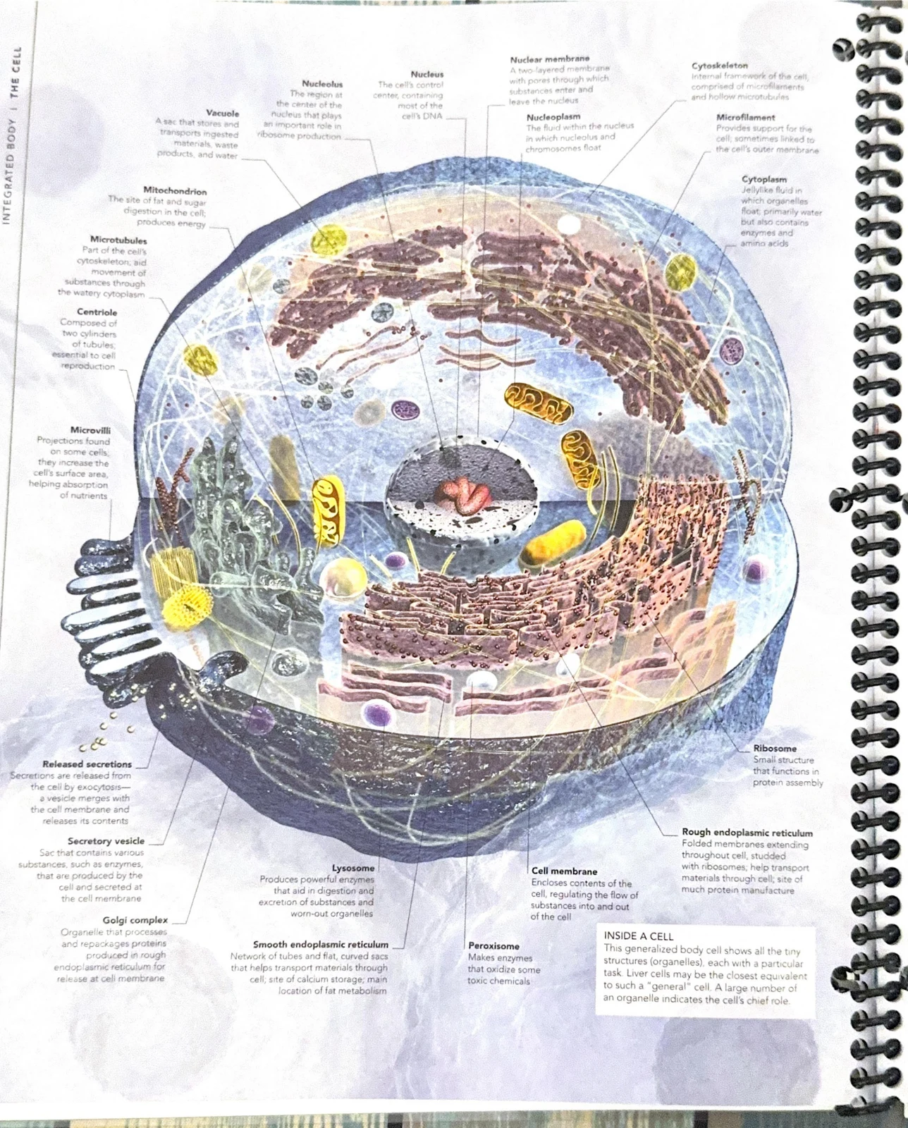
A cell is not merely a bag of chemicals; it is a highly organized environment containing specialized structures called organelles. These organelles are held within the cytoplasm, a cellular fluid that is primarily water but also contains essential enzymes and amino acids.
This diagram illustrates the structure and organelles of a generalized animal cell, including the nucleus, mitochondria, endoplasmic reticulum, and Golgi apparatus.
Key Organelles and Their Roles
Each organelle has a characteristic shape, size, and function, ensuring that the cell's internal processes are compartmentalized and efficient.
The Nucleus: Often described as the cell's control center, the nucleus houses the majority of the cell's DNA. This genetic material contains the instructions for how the cell grows, develops, and functions.
Mitochondrion: Known as the "powerhouse" of the cell, the mitochondrion is responsible for energy production. Its inner membrane is uniquely folded into partitions to increase the surface area available for releasing energy from sugars and fats. Interestingly, mitochondria possess their own unique, circular DNA (mtDNA) that is inherited exclusively from the mother.
Endoplasmic Reticulum (ER): This is a network of highly folded membranes that creates a labyrinthine space within the cell. The rough ER is studded with ribosomes, making it a primary site for protein manufacture and transport.
Ribosomes: These are small structures that function as the primary sites for protein assembly.
Golgi Complex: Acting as the cell's "post office," the Golgi complex consists of a stack of flattened membranous sacs. It receives proteins from the ER, modifies them, and repackages them for transport.
Student Tip: The "Factory" Analogy
To remember organelle functions, think of a factory: The Nucleus is the CEO's office (blueprints/DNA); the Ribosomes are the workers; the Rough ER is the assembly line; the Golgi Complex is the shipping and receiving department; and the Mitochondria is the power plant.
The Gatekeeper: Cell Membrane and Transport Mechanisms
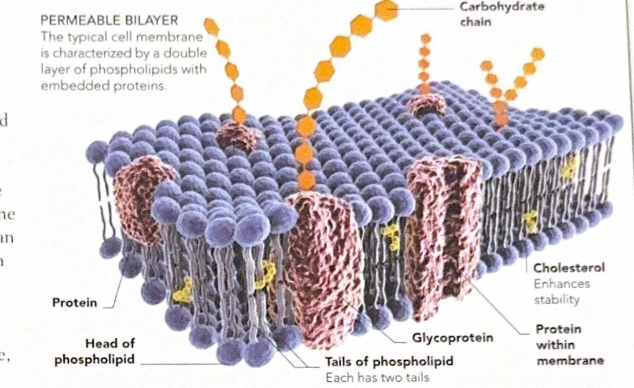
The cell is separated from its environment by the cell membrane. This membrane is not a solid wall but a dynamic, double layer of phospholipids known as a phospholipid bilayer.
Diagram of a permeable bilayer cell membrane, illustrating phospholipids, embedded proteins, glycoproteins, and carbohydrate chains.
Each phospholipid molecule consists of a water-loving (hydrophilic) head and two water-fearing (hydrophobic) tails. In the bilayer, the heads face the watery environments inside and outside the cell, while the tails hide in the middle. Interspersed within this layer are proteins and carbohydrate chains that allow the cell to recognize other cells.
How Materials Move
The membrane regulates the movement of substances through three primary processes:
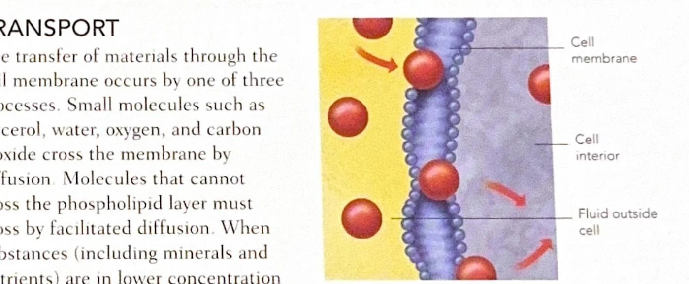
Diffusion: Small, uncharged molecules like oxygen, carbon dioxide, water, and glycerol move naturally from an area of high concentration to an area of low concentration.
Diagram illustrating the transport of molecules across a cell membrane via diffusion and facilitated diffusion.
Facilitated Diffusion: Molecules that are too large or cannot pass through the lipid layer must use a "helper." A carrier protein binds to a specific molecule (like glucose) on the outside of the cell, changes shape, and ejects it into the cell.
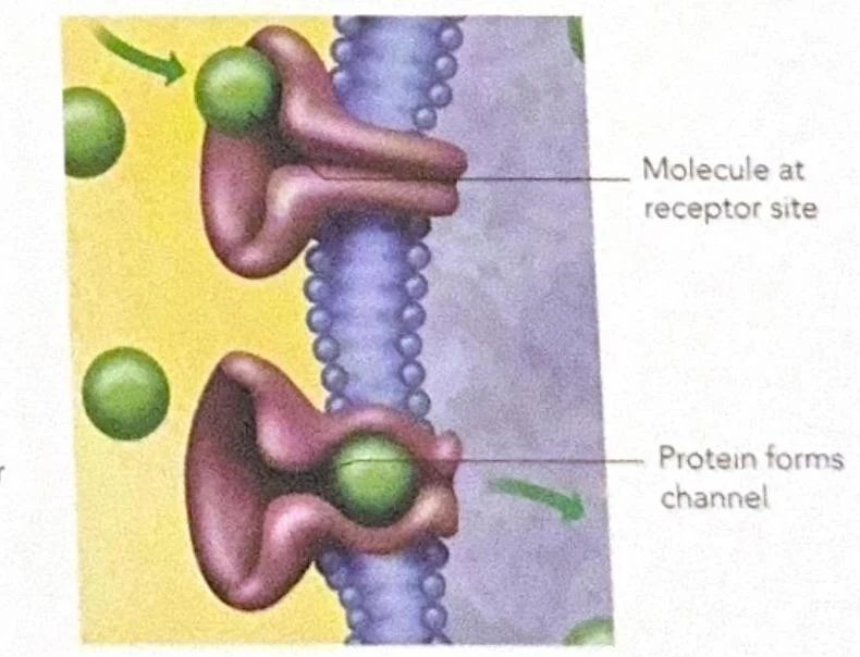
Active Transport: When a cell needs to move substances (like minerals or nutrients) against a concentration gradient—from a low concentration to a high concentration—it must use energy. This often involves a molecule binding to a receptor, triggering a protein to form a channel for transport.
Diagram illustrating a molecule binding to a receptor site on a cell membrane, with the protein forming a channel.
From Cells to Tissues: The Four Primary Types
When a group of similar cells work together to perform a specialized function, they form a tissue. While the human body contains over 200 types of specialized cells, they are categorized into four primary tissue types: epithelial, connective, muscle, and nervous.
1. Epithelial Tissue
Epithelial cells form the skin and line the internal organs and hollow cavities. Because these cells are often subject to physical wear and tear (such as the lining of the intestinal tract), they typically have very rapid division rates to replace damaged cells.
2. Connective Tissue
Connective tissue is diverse, serving to support, bind, and protect body parts. It is categorized based on its structure:
Loose Connective Tissue: Consists of cells like fibroblasts embedded in a matrix of fibers, such as elastin and collagen.
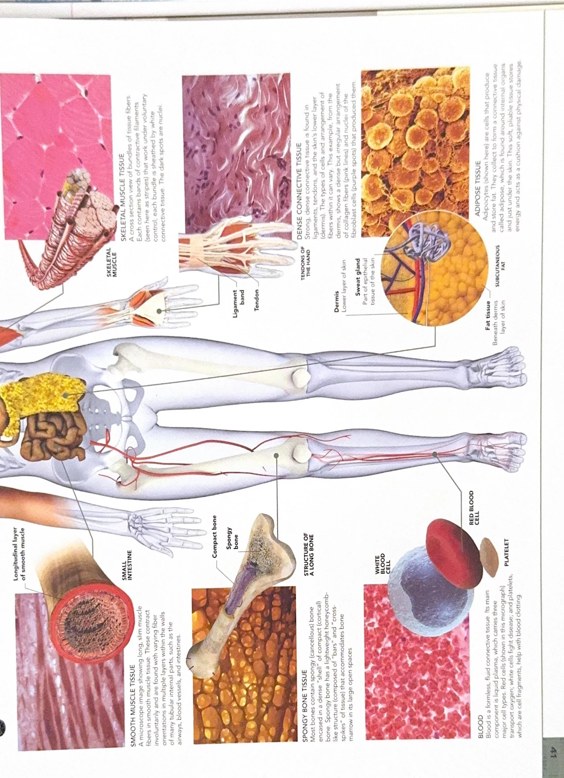
Dense Connective Tissue: Contains strong, thick bundles of collagen. This is found in high-stress areas like tendons and ligaments.
This illustration displays various types of human tissues, including skeletal muscle, dense connective tissue, adipose tissue, smooth muscle, spongy bone, and blood cells.
Specialized Connective Tissues: This category includes several distinct forms:
Adipose Tissue: Fat-storing cells called adipocytes that store energy as lipids.
Cartilage: Includes hyaline (tough yet flexible) and elastic cartilage (light and bendy).
Bone: A rigid structural tissue.
Blood: A unique fluid connective tissue containing erythrocytes (red blood cells), leukocytes (white blood cells), and platelets.
3. Muscle Tissue
Muscle tissue is specialized for contraction and is responsible for all body movement.
4. Nervous Tissue
Nervous tissue is designed for communication. It is composed of neurons, which conduct messages, and neuroglia, which are support cells. For example, astrocytes are a type of neuroglia that provide essential nutrients to neurons.
Tissues are composed of specialized cells and are classified into four primary types: epithelial, connective, muscle, and nervous tissue.
Case Study: The Liver and Specialized Cells
To see how these tissues interact, consider the liver. The liver is a complex organ composed of various tissues, including epithelial (hepatocytes), connective, and nervous tissues.
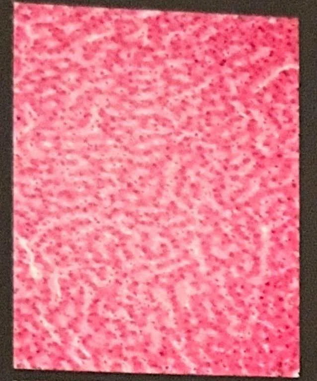
Microscopic view of liver tissue showing hepatocytes and sinusoids.
In the liver, hepatocytes (liver cells) are arranged in branching sheets called laminae. Between these cells are hepatic sinusoids, which are highly porous blood vessels that allow for the efficient exchange of oxygen and nutrients. A specialized type of white blood cell called a Kupffer cell (a hepatic macrophage) resides here to engulf and digest old blood cells and debris.
Cells also exhibit unique shapes (morphologies) to suit their specific jobs:
Erythrocytes (Red Blood Cells): These have a biconcave (double-dished) shape, which maximizes their surface area for rapid oxygen absorption.
Cone Cells: Found in the retina of the eye, these are specialized light-sensitive neurons responsible for color perception.
Adipocytes: These are bulky cells "crammed" with fat droplets to store energy.
The Genetic Blueprint: DNA and Protein Synthesis
The instructions for building and operating these cells are contained within DNA (deoxyribonucleic acid). In almost all human cells, DNA is organized into 46 X-shaped chromosomes located in the nucleus.
DNA Structure and the Genome
DNA is a double helix—a twisted ladder structure. The "rungs" of this ladder are made of four chemical bases: Adenine (A), Thymine (T), Guanine (G), and Cytosine (C). These bases follow strict pairing rules: A always pairs with T, and G always pairs with C.
The complete set of genetic instructions for a living organism is called the genome. The human genome contains an estimated 20,000 genes. A gene is a specific unit of DNA that provides the code to construct one protein.
From Gene to Protein: Transcription and Translation
The process of turning genetic code into a functional protein happens in two main stages:
Transcription: This occurs in the nucleus. The DNA sequence is copied into a messenger molecule called mRNA (messenger RNA).
Translation: The mRNA travels out of the nucleus to the ribosomes. Here, the mRNA acts as a template. Molecules called tRNA (transfer RNA) bring specific amino acids to the ribosome, matching them to the mRNA's "triplet codons". The amino acids are then linked together to form a protein chain.
Edge Case: Non-coding DNA
Students often assume all DNA codes for something. However, a significant portion of the genome is "non-coding" or "junk" DNA. While it doesn't code for proteins, it plays a vital role in regulating how and when genes are turned on or off.
Cellular Differentiation and Stem Cells
How does a single fertilized egg become a complex body with skin, bone, and brain cells? The answer lies in cellular differentiation.
All cells begin as stem cells—undifferentiated cells that have the potential to become specialized.
Embryonic Stem Cells: Found in the early embryo, these can differentiate into any of the 200+ cell types in the body.
Adult Stem Cells: Found in specific tissues (like bone marrow), these help with ongoing maintenance and repair.
As cells develop, certain genes are "switched on" (expressed) while others are "switched off". This process is influenced by the cell's environment and can even be regulated by epigenetic changes—chemical modifications to DNA that affect gene expression without changing the underlying genetic code.
Keep reading for free.
Create an account to finish this reading and make it part of your study routine.