Cellular Adaptation and Injury: Mechanisms of Cellular Response
Explore how cells adapt to stress through atrophy, hypertrophy, hyperplasia, metaplasia, and dysplasia. Understand the progression from reversible to irreversible cell injury, leading to necrosis and apoptosis.
Cells are dynamic entities that exist in a state of constant equilibrium with their environment. To maintain homeostasis, cells must possess the ability to sense changes in their surroundings and respond accordingly. When a cell encounters physiological stress or pathological stimuli, it undergoes a series of adjustments to survive. This article explores the spectrum of cellular responses, ranging from beneficial adaptations to the point of no return: irreversible cell injury.
Cellular Adaptation: The Survival Response
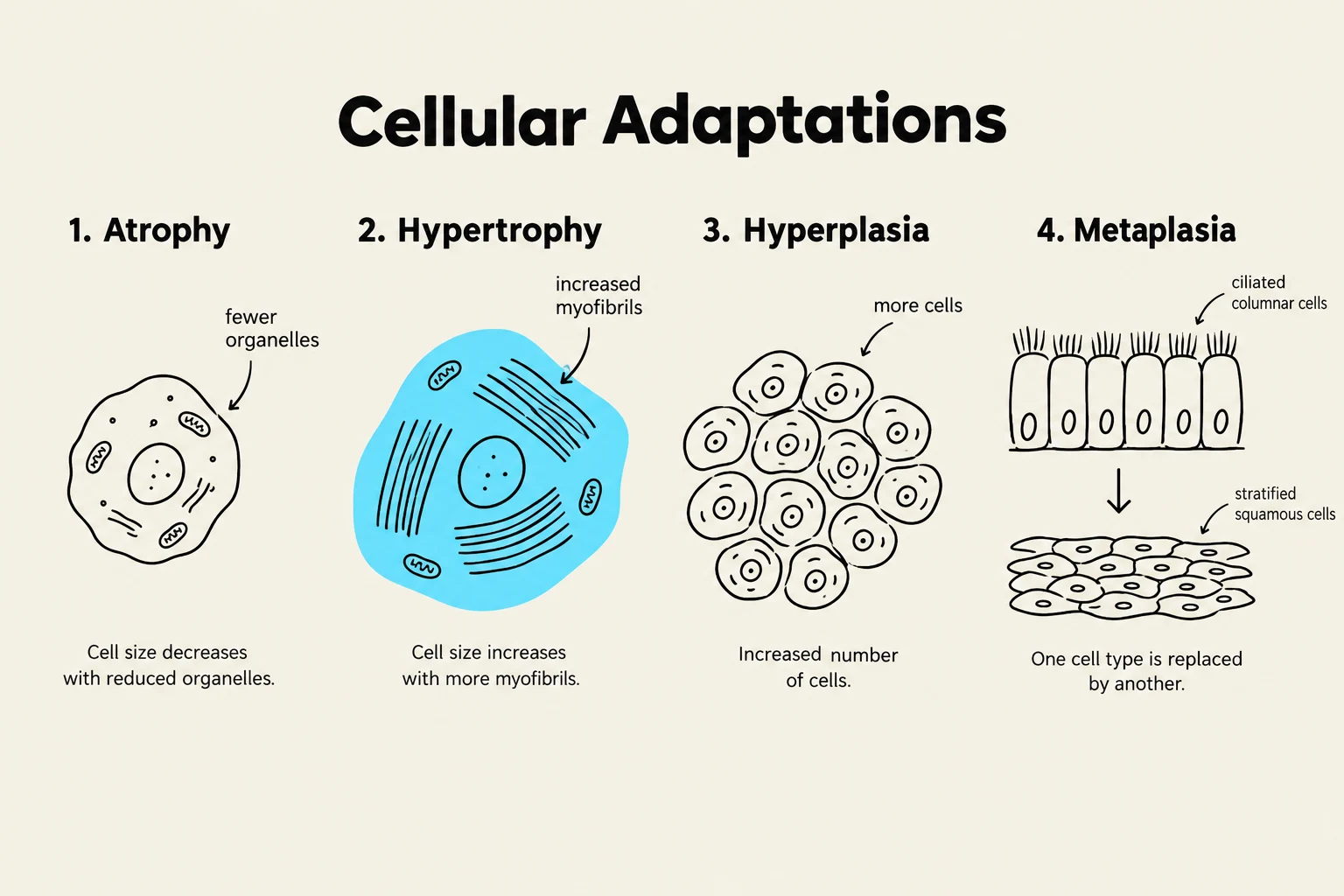
This diagram illustrates four types of cellular adaptations: atrophy, hypertrophy, hyperplasia, and metaplasia, with accompanying visual representations and brief explanations.
When cells are subjected to stress, they often undergo structural or functional changes to achieve a new steady state. These changes are known as cellular adaptations. While these processes allow the cell to persist in a changing environment, they are not "normal" in the sense of baseline homeostasis; rather, they are compensatory mechanisms.
Atrophy
Atrophy is a decrease in the size of the cell, which subsequently leads to a decrease in the size of the organ. This can occur due to several factors, such as decreased workload (disuse atrophy), loss of innervation, diminished blood supply (ischemia), inadequate nutrition, or loss of endocrine stimulation. At the molecular level, atrophy is driven by an imbalance between protein synthesis and protein degradation, often involving the ubiquitin-proteasome pathway.
Hypertrophy
In contrast to atrophy, hypertrophy is an increase in the size of cells, resulting in an increase in the size of the organ. This typically occurs in cells that have a limited capacity to divide, such as cardiac muscle or skeletal muscle. Hypertrophy is driven by increased protein synthesis and the production of more cellular components (like myofibrils) to handle increased functional demands.
Hyperplasia
Hyperplasia refers to an increase in the number of cells in an organ or tissue. Unlike hypertrophy, which focuses on cell size, hyperplasia is a proliferative response. It can be physiological (such as the proliferation of the glandular epithelium in the female breast during puberty) or pathological (such as the excessive growth of the uterine lining in endometrial hyperplasia).
Metaplasia
Metaplasia is a reversible change in which one adult cell type (epithelial or mesenchymal) is replaced by another adult cell type. This is often a protective mechanism against chronic irritation. For example, in the respiratory tract of a chronic smoker, the normal ciliated columnar epithelium may be replaced by stratified squamous epithelium, which is more rugged and better able to survive the irritation of smoke. However, this comes at a cost: the loss of the specialized functions of the original cells, such as mucus secretion and ciliary clearance.
Dysplasia
Dysplasia is characterized by disordered growth and maturation of an epithelium. It involves a loss of cellular uniformity and architectural orientation. While dysplasia is not "cancer" per se, it is often considered a pre-neoplastic change. If the stimulus causing the dysplasia is removed, the tissue may return to normal; however, if the stimulus persists, dysplasia can progress to malignancy.
Exam Tip: Distinguish between metaplasia and dysplasia. Metaplasia is a replacement of one mature cell type with another mature cell type (a controlled adaptation). Dysplasia is a disorganized, disordered growth pattern that is often a precursor to cancer.
Cellular Injury: Crossing the Threshold
When the limits of adaptive responses are exceeded, or when the stress is inherently harmful (such as toxins, hypoxia, or physical trauma), cellular injury occurs. Injury is categorized into two stages: reversible and irreversible.
Reversible Cell Injury
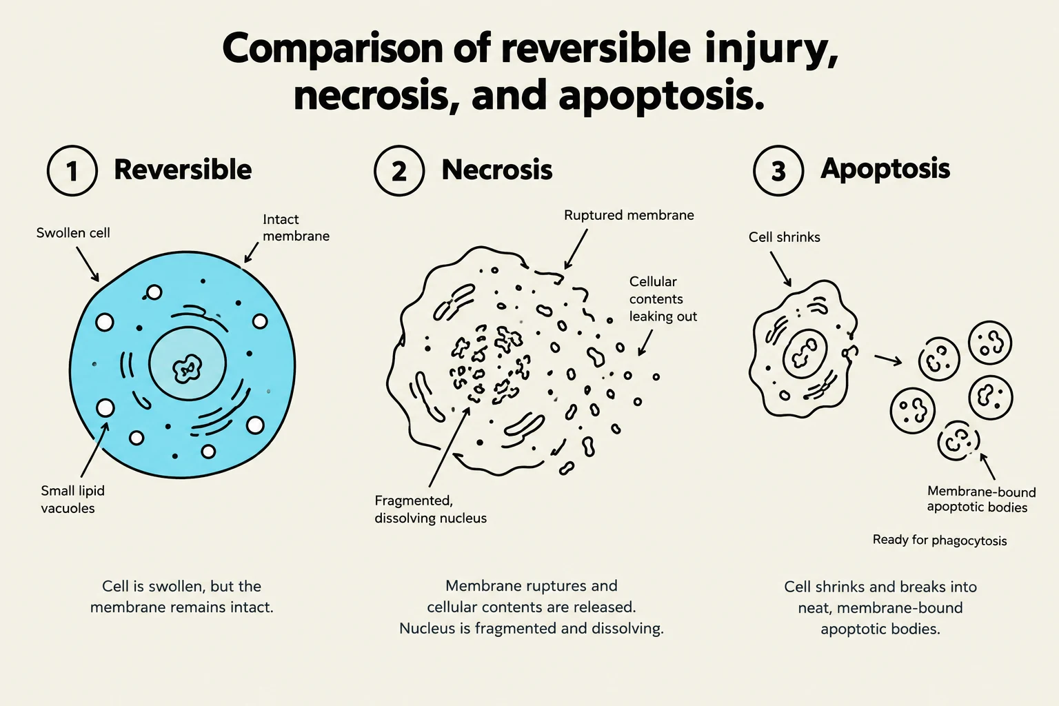
In the early stages of injury, the changes are still capable of being undone if the stimulus is removed. The hallmark of reversible injury is cellular swelling (oncosis) and fatty change.
Cellular Swelling: This occurs when the cell cannot maintain ionic and fluid homeostasis, often due to the failure of energy-dependent membrane pumps (like the Na+/K+-ATPase pump). When ATP levels drop, sodium accumulates inside the cell, drawing water in via osmosis.
Fatty Change: In organs involved in lipid metabolism (like the liver), injury may manifest as the appearance of lipid vacuoles within the cytoplasm.
Irreversible Cell Injury and Cell Death
This diagram illustrates and compares the cellular changes that occur during reversible injury, necrosis, and apoptosis.
If the injury persists or is severe enough, the cell passes a "point of no return" and undergoes irreversible injury, leading to cell death. The two primary patterns of cell death are necrosis and apoptosis.
Necrosis
Necrosis is always a pathological process. It is characterized by cell swelling, protein denaturation, and enzymatic digestion of the cell. A critical feature of necrosis is the loss of membrane integrity, which allows cellular contents to leak into the surrounding tissue, often triggering an inflammatory response. Common patterns of necrosis include:
Coagulative necrosis: Seen in infarcts (ischemia) in most organs except the brain.
Liquefactive necrosis: Typical of focal bacterial infections and brain infarcts.
Caseous necrosis: Characteristic of tuberculosis (resembling "cheese").
Apoptosis
Apoptosis is programmed cell death, which can be either physiological (e.g., during embryogenesis or the shedding of the uterine lining) or pathological (e.g., when a cell's DNA is irreparably damaged). Unlike necrosis, apoptosis does not typically elicit an inflammatory response because the cell fragments into "apoptotic bodies" that are neatly phagocytosed by neighboring cells without leaking contents.
Summary of Mechanisms
The transition from adaptation to death is fundamentally a failure of energy production and membrane stability.
Feature
Reversible Injury
Irreversible Injury (Necrosis)
Cell Size
Swelling
Swelling and rupture
Nucleus
Normal or pyknosis
Fragmentation (karyorrhexis/karyolysis)
Plasma Membrane
Blebbing, loss of microvilli
Extensive damage/rupture
Mitochondria
Swelling
Severe swelling, large densities
The progression of cellular damage can be summarized by the following logic: