


To understand the complexity of the human body, one must first look at its most fundamental components. The body is organized into a hierarchy: individual cells form tissues, tissues form organs, and organs work together within systems to maintain life. This article explores the anatomy of the cell, the four primary tissue types, and the protective structures of the integumentary system.
Anatomy of the Cell
The cell is the basic functional unit of life. While cells vary significantly in shape and function depending on their location in the body, they share several critical organelles that allow them to perform life-sustaining processes.
- Nucleus: Often referred to as the control center, the nucleus houses the cell's genetic material (DNA) and coordinates activities such as growth and reproduction.
- Cytoplasm: This is the jelly-like substance that fills the interior of the cell, surrounding the organelles and providing a medium for chemical reactions.
- Cell Membrane: A semi-permeable barrier that surrounds the cell, regulating the passage of substances (such as nutrients and waste) in and out of the cell.
- Mitochondria: Known as the "powerhouse" of the cell, these organelles are the primary site of energy production through cellular respiration.
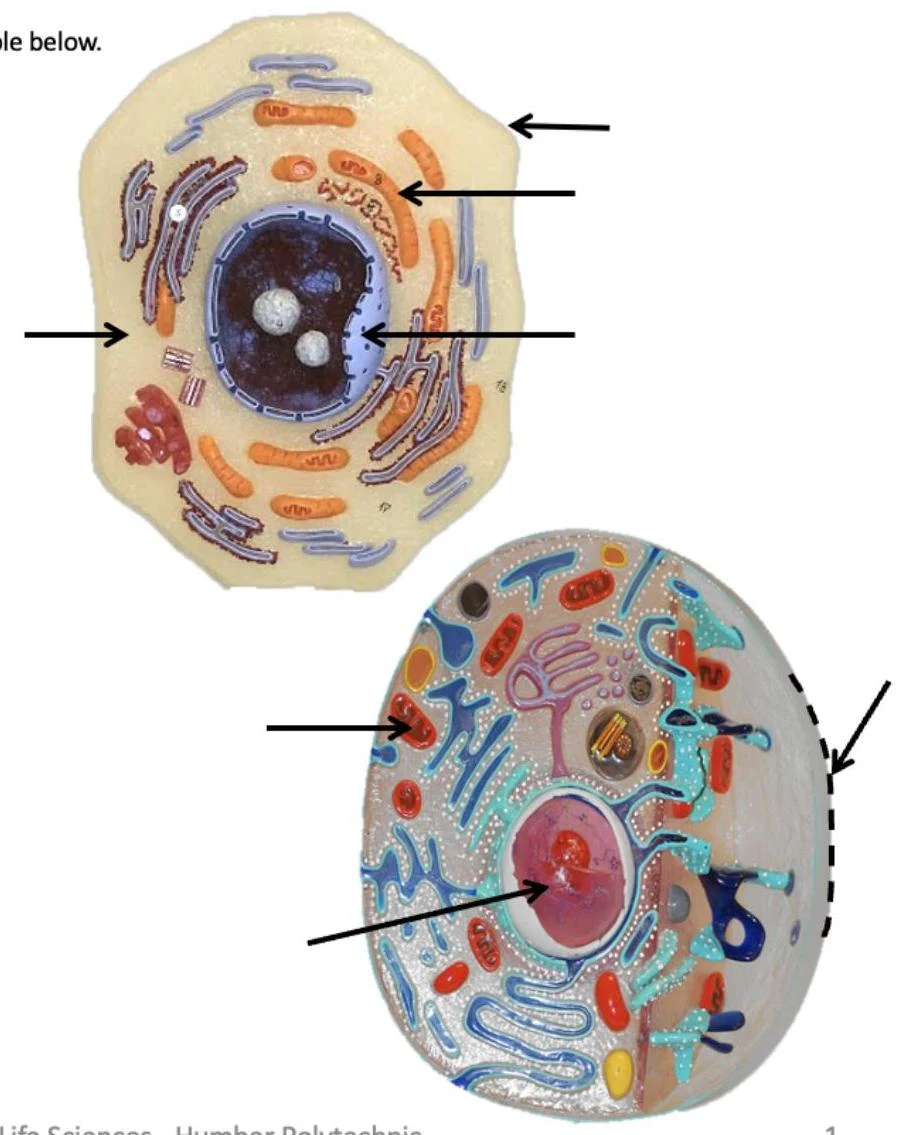
Exam Tip: In lab "Bell Ringers," you may be asked to identify these organelles on a 3D model. Be sure you can distinguish the nucleus from the surrounding cytoplasm and identify the membrane as the outermost boundary.
Epithelial Tissue
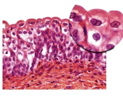
Epithelial tissues cover body surfaces, line internal cavities, and form glands. They are classified based on the number of cell layers and the shape of the cells.


Keep reading for free.
Create an account to finish this reading and make it part of your study routine.
Continue reading freeAlready have an account? Log in
Updated