Explores the fundamental structure of cells, including the plasma membrane, nucleus, and cytoplasm, and details the mechanisms of membrane transport, such as passive, active, and vesicular transport.
The human body is a staggering collection of biological complexity, composed of approximately 50 to 100 trillion cells. While these cells vary immensely in size and shape—ranging from microscopic structures to nerve cells that can reach a yard in length—they all share a fundamental biological principle: the cell is the basic structural and functional unit of life. The collective activities of these cells dictate the overall function of the entire organism.
Cell Structure and Function
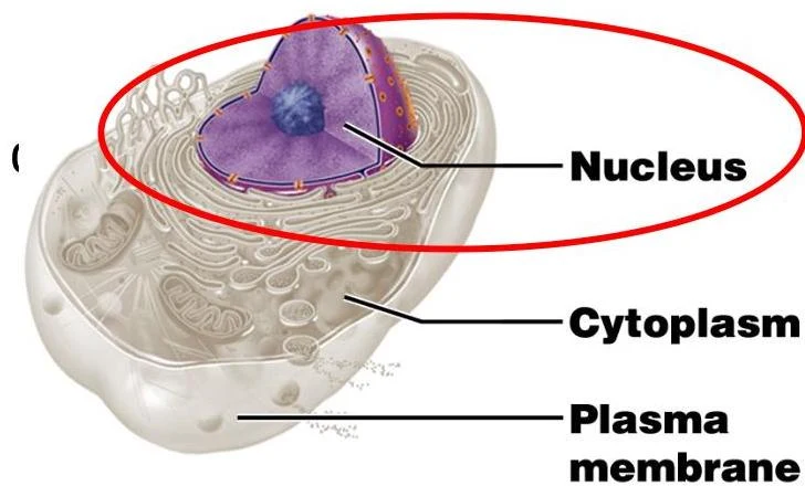
A cell's biochemical activities are directly dictated by its anatomy; essentially, its structure determines its physiology. While cells are specialized to perform diverse tasks such as metabolizing nutrients, digesting food, disposing of waste, growing, and responding to stimuli, a generalized cell can be divided into three main regions:
The Plasma Membrane: The outer boundary that regulates interaction with the environment.
The Nucleus: The control center containing genetic material.
The Cytoplasm: The internal region containing the cell's metabolic machinery.
Diagram of a cell nucleus, cytoplasm, and plasma membrane.
The Plasma Membrane and the Fluid-Mosaic Model
The plasma membrane acts as a transparent barrier that contains the cell's contents and separates them from the surrounding environment. To describe its complex nature, scientists use the fluid-mosaic model. This model describes the membrane as a "mosaic" composed of various lipid, protein, and carbohydrate molecules.
The Phospholipid Bilayer
The foundation of the membrane is the phospholipid bilayer. Phospholipids are unique molecules with:
Hydrophilic ("water-loving") polar heads: These orient themselves outward toward the inner and outer surfaces of the membrane.
Hydrophobic ("water-fearing") nonpolar tails: These cluster together to form the interior center of the membrane.
Diagram illustrating the structure of a cell membrane, highlighting the phospholipid bilayer, integral and peripheral membrane proteins, glycoproteins, glycolipids, and cholesterol.
Membrane Proteins
Embedded within or attached to this bilayer are various proteins that facilitate specific cellular functions:
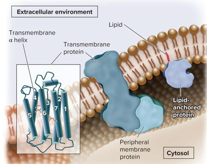
Transmembrane (Integral) Proteins: These have one or more regions physically embedded within the phospholipid bilayer.
Lipid-anchored Proteins: These are attached to the membrane via a lipid molecule that is covalently bonded to an amino acid side chain, with the lipid tails inserted into the membrane.
Diagram illustrating the structure of a cell membrane, showing transmembrane proteins, peripheral membrane proteins, and lipid-anchored proteins within the extracellular environment and cytosol.
Selective Permeability and Membrane Transport
The plasma membrane is selectively permeable, meaning it regulates what enters and exits the cell. This allows essential nutrients to enter while preventing undesirable substances from passing through.
To understand how substances move, we must distinguish between the fluids surrounding the cell. The intracellular fluid (including the nucleoplasm and cytosol) is the solution of gases, nutrients, and salts dissolved in water inside the cell. The interstitial fluid is the fluid located on the exterior of the cell.
Passive Transport
Passive transport requires no input of cellular energy because substances move "down" or "with" their concentration gradient (from high to low concentration).
Simple Diffusion: Solutes move directly through the phospholipid bilayer. This is common for small, lipid-soluble materials.
Osmosis: A specific type of simple diffusion involving the movement of water, often through specialized channel proteins called aquaporins.
Facilitated Diffusion: Substances move through the membrane with the aid of a transport protein. This is the primary pathway for organic molecules like sugars, amino acids, and nucleotides.
Exam Tip: Transporter Types in Facilitated Diffusion
Uniporter: Transports a single molecule or ion.
Symporter (Cotransporter): Transports two or more substances in the same direction.
Antiporter: Transports two or more substances in opposite directions.
Active Transport and Vesicular Transport
When substances must move against a concentration gradient (from low to high concentration), the cell must use energy, typically in the form of ATP.
Solute Pumping (Active Transport): Protein carriers called solute pumps use ATP to move ions and molecules. A classic example is the Na+/K+-ATPase pump, which pumps three Na+ ions out of the cell and two K+ ions into the cell to maintain electrochemical gradients necessary for nerve impulses.
Vesicular Transport: For much larger materials, the cell uses membranes to "package" substances.
Exocytosis: Vesicles migrate to and fuse with the plasma membrane to release contents outside the cell.
Endocytosis: The cell engulfs extracellular substances by enclosing them in a vesicle. This includes phagocytosis ("cell eating"), pinocytosis ("cell drinking"), and receptor-mediated endocytosis.
The Nucleus and Cytoplasm: The Cell's Internal Machinery
The Nucleus
The nucleus serves as the cell's control center, housing the DNA required for protein synthesis and reproduction. It consists of the nuclear envelope (a double membrane with pores), the nucleolus (the site of ribosome assembly), and chromatin (DNA wound around proteins).
The Cytoplasm and Organelles
The cytoplasm is composed of the cytosol (the fluid), inclusions (stored nutrients), and organelles (the metabolic machinery). Key organelles include:
Mitochondria: The "powerhouses" of the cell that synthesize ATP.
Ribosomes: The sites of protein synthesis.
Endoplasmic Reticulum (ER): The Rough ER (studded with ribosomes) synthesizes and transports proteins, while the Smooth ER functions in lipid metabolism and detoxification.
Golgi Apparatus: Modifies, packages, and sorts proteins arriving from the ER.
Lysosomes and Peroxisomes: Lysosomes contain digestive enzymes to break down waste, while peroxisomes use oxidase enzymes to detoxify harmful substances like alcohol.
Cytoskeleton: A network of microtubules, intermediate filaments, and actin filaments that provides structural support and enables movement.
Cellular Movement
Cells can move using specialized structures derived from the cytoskeleton:
Pseudopodia: Temporary, finger-like projections used by amoebas for movement and engulfing particles.
Flagella: Long, whip-like structures that generate movement (e.g., in sperm cells).
Cilia: Shorter structures that often cover the cell surface to move the cell or surrounding fluids.
Keep reading for free.
Create an account to finish this reading and make it part of your study routine.