Cell Membrane Structure, Transport, and Electrical Signaling
Explores the fluid mosaic model of the cell membrane, including its lipid and protein components, selective permeability, and mechanisms of passive and active transport. It also covers ion channels, membrane potentials, the Nernst equation, and the generation and propagation of action potentials.
The cell membrane is far more than a simple container; it is a dynamic, selective barrier that makes every other physiological process—from nutrient uptake to electrical signaling—possible. By controlling what enters and exits the cell, the membrane establishes the chemical gradients necessary for life and serves as the primary interface for communication between cells.
The Fluid Mosaic Model and Membrane Structure
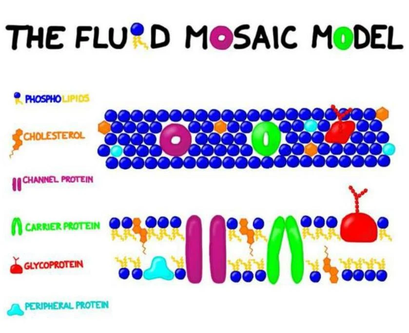
The modern understanding of the cell membrane is described by the fluid mosaic model. This model depicts the membrane as a flexible, shifting sea of lipids in which various proteins are embedded, much like tiles in a mosaic.
The fluid mosaic model illustrates the structure of cell membranes, depicting phospholipids, cholesterol, and various proteins like channel proteins, carrier proteins, glycoproteins, and peripheral proteins.
The Lipid Component
The foundation of the membrane is the phospholipid bilayer. Each phospholipid molecule consists of a phosphorylated glycerol head (hydrophilic) and two fatty acid tails (hydrophobic). This arrangement creates a barrier that is naturally impermeable to most charged or polar substances. Additionally, glycolipids are present on the surface, playing critical roles in cell recognition and immune responses.
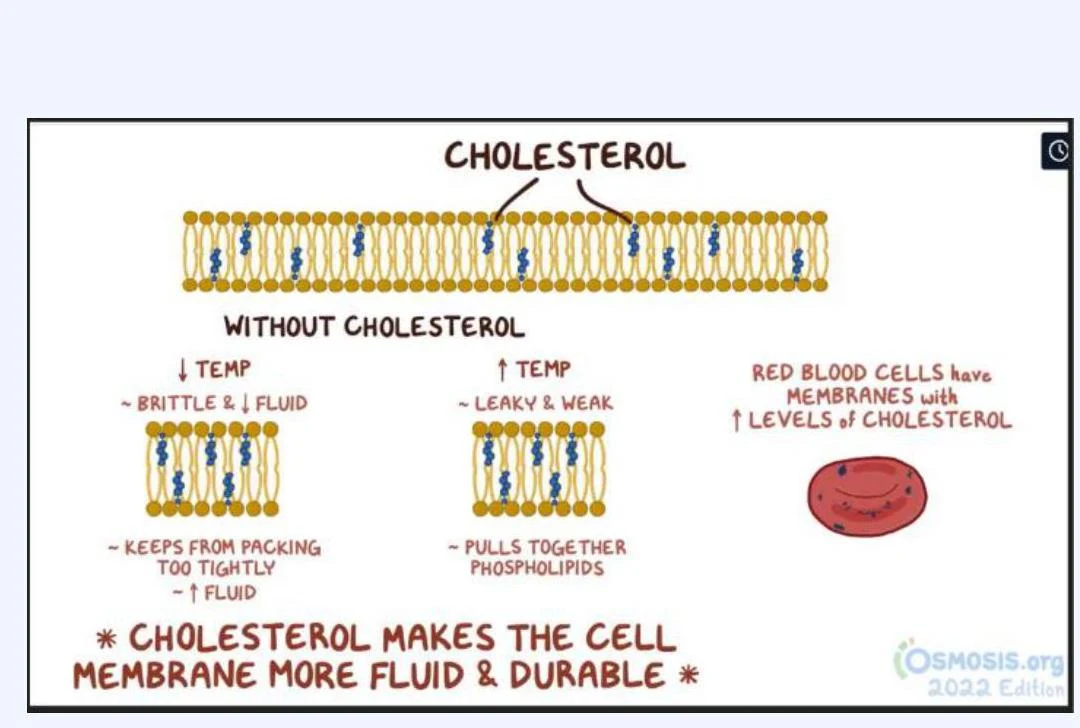
Cholesterol is embedded between these phospholipids to modulate membrane fluidity:
At low temperatures: It prevents phospholipids from packing too tightly, maintaining fluidity and preventing the membrane from becoming brittle.
At high temperatures: It pulls molecules together to decrease space, preventing leaks and maintaining structural integrity.
This illustration explains the role of cholesterol in cell membranes. Without cholesterol, membranes can become brittle at low temperatures and leaky at high temperatures. Cholesterol helps maintain membrane fluidity and durability across a range of temperatures. Red blood cells are shown to have membranes with increased levels of cholesterol.
The Protein Component
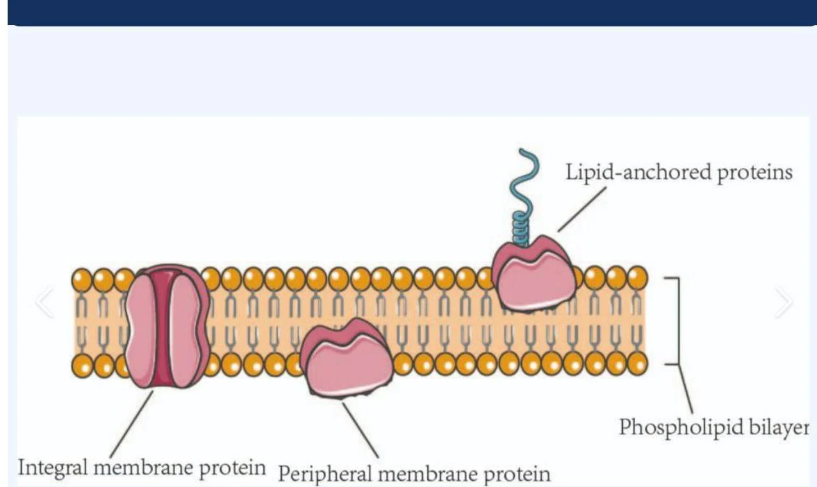
Proteins provide the membrane with its functional diversity, categorized by how they associate with the bilayer:
Integral proteins: These are embedded in or anchored to the membrane via hydrophobic interactions. Some are "transmembrane" proteins that span the entire bilayer.
Peripheral proteins: These are loosely attached to either the extracellular (EC) or intracellular (IC) sides through electrostatic interactions.
Diagram illustrating the structure of a cell membrane, showing integral membrane proteins, peripheral membrane proteins, and lipid-anchored proteins embedded within a phospholipid bilayer.
Selective Permeability and Membrane Transport
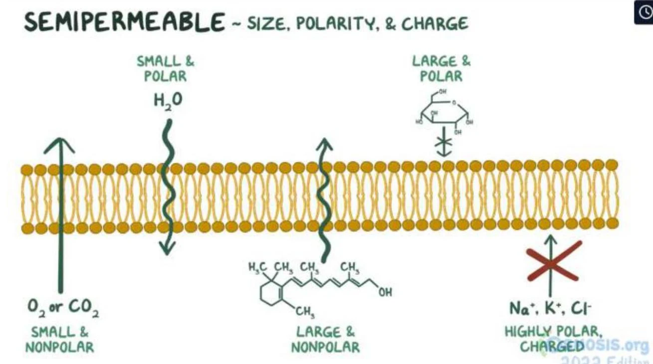
The membrane's composition dictates selective permeability—the rule that permeability rises with lipid solubility and falls with size and charge. Small, nonpolar molecules like oxygen and carbon dioxide can dissolve directly into the lipid core and cross easily. In contrast, highly polar or charged ions (such as Na+, K+, and Ca2+) cannot cross the bilayer on their own and require specialized proteins.
This diagram illustrates how the semipermeable membrane's properties (size, polarity, and charge) affect the passage of different molecules, such as water, oxygen, carbon dioxide, glucose, and ions.
Passive Transport
Passive transport moves solutes down their electrochemical gradient without the expenditure of metabolic energy (ATP).
Simple Diffusion: Solutes move directly through the lipid core or through an open channel. This process is driven by thermal motion and follows Fick's Law: flux is proportional to the steepness of the concentration gradient, surface area, and the diffusion coefficient.
Facilitated Diffusion: Solutes require a transport protein (a carrier or a channel) to cross. While channels allow for rapid flux, carrier proteins exhibit "saturation" (a transport maximum) because they must bind the solute to function.
Active Transport
Active transport moves solutes against their electrochemical gradient, which requires energy.
Primary Active Transport: Uses energy derived directly from ATP hydrolysis. A classic example is the Na+/K+ ATPase, which pumps three Na+ ions out and two K+ ions in per ATP molecule hydrolyzed. This pump is "electrogenic," meaning it contributes directly to the negative resting potential.
Secondary Active Transport: Does not use ATP directly. Instead, it uses the energy stored in the gradient of a second solute (usually Na+) created by a primary active transporter. This occurs via symport (moving two solutes in the same direction, like glucose via SGLT) or antiport (moving solutes in opposite directions, like the Na+/Ca2+ exchanger).
Ion Channels and Gating Mechanisms
Ion channels are specialized proteins that act as "doors" for specific ions. They are classified by their gating mechanisms—how they open and close:
Leak channels: Always open; they set the baseline permeability that determines the resting membrane potential.
Voltage-gated channels: Open in response to changes in the membrane potential; these are essential for generating action potentials.
Ligand-gated channels: Open when a specific chemical messenger (a ligand) binds to the receptor.
Mechanically gated channels: Open in response to physical deformation, such as stretch or pressure.
Chemical Signaling and GPCRs
Cellular communication often involves ligands binding to receptors on the cell surface. One of the most significant families of receptors is the G-protein coupled receptors (GPCRs). When a ligand binds to a GPCR, it activates an intracellular G-protein, which then triggers a cascade of second messengers (like cAMP or Ca2+).
These cascades provide amplification: a single ligand binding to one receptor can activate millions of downstream enzymes, allowing cells to respond to incredibly low concentrations of hormones or neurotransmitters. To prevent overstimulation, signals are terminated through messenger breakdown, G-protein inactivation, or receptor desensitization.
Membrane Potentials and the Nernst Equation
The membrane potential (Vm) is the difference in electrical charge between the inside and outside of the cell. This potential acts like a battery, providing energy for transport and enabling electrical signaling.
The Resting Membrane Potential
At rest, a typical neuron has a Vm of approximately −70 mV. This negative value is primarily due to the high permeability of the membrane to K+ via leak channels. While the equilibrium potential for K+ (EK) is roughly −91 mV, the actual resting potential is slightly more positive because of small leaks of Na+ into the cell.
The Nernst Equation
To find the equilibrium potential (Eion) for a single ion—the voltage at which that ion would have no net movement—we use the Nernst equation:
Eion=61 mV×log10([Ion]inside[Ion]outside)(Note: The 61 mV term is a constant for body temperature; for divalent ions like Ca2+, the value is divided by 2).
Exam Tip: The Nernst equation tells you the equilibrium potential for one ion in isolation. It does not predict the actual resting membrane potential, which is a weighted average of all permeable ions.
The Action Potential
The action potential is an "all-or-none" electrical signal that propagates along excitable cells like neurons. It is driven by the rapid opening and closing of voltage-gated channels:
Threshold: A stimulus must depolarize the membrane to about −55 mV to trigger the self-reinforcing opening of voltage-gated Na+ channels.
Depolarization:Na+ rushes into the cell, driving the Vm toward the Na+ equilibrium potential (+60 mV).
Repolarization:Na+ channels inactivate, and slower voltage-gated K+ channels open, allowing K+ to exit the cell.
Hyperpolarization:K+ channels close slowly, causing the Vm to undershoot the resting potential.
Refractory Periods
The timing of these channels creates two distinct refractory periods:
Absolute Refractory Period: From threshold through most of repolarization, Na+ inactivation gates are closed. No stimulus, regardless of strength, can trigger another action potential.
Relative Refractory Period: During late repolarization, some Na+ channels have reset, but K+ conductance remains high. A stronger-than-normal stimulus is required to fire.
Clinical Connection: In diseases like Multiple Sclerosis, demyelination slows conduction velocity because the signal can no longer "jump" between nodes of Ranvier via saltatory conduction.
Keep reading for free.
Create an account to finish this reading and make it part of your study routine.