Explores the dynamic structure of the cell membrane, including the phospholipid bilayer, membrane fluidity, proteins, selective permeability, and cell junctions. Discusses visualization techniques and the role of the extracellular matrix.
The cell membrane is far more than just a simple container; it is a dynamic, flexible, and highly organized boundary that defines the very essence of life. Without the ability to form a membrane, a cell could not establish a unique identity or separate its internal metabolic processes from the external environment. By creating this delimitation, the membrane allows for compartmentalization, enabling specialized activities—such as fatty acid synthesis in one area and oxidation in another—to occur simultaneously without interference.
The Phospholipid Bilayer: The Foundation of Life
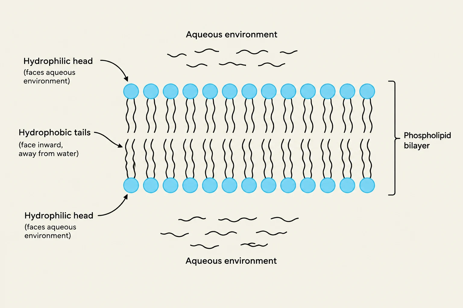
At the heart of every membrane is the phospholipid bilayer. Phospholipids are amphipathic molecules, meaning they possess two distinct chemical personalities: a hydrophilic (water-loving) head and two hydrophobic (water-fearing) tails.
In the aqueous environment of a living organism, these molecules spontaneously assemble into a bilayer. The hydrophilic heads face outward toward the water (both inside and outside the cell), while the hydrophobic tails hide in the interior, shielded from water. This arrangement is driven by hydrophobic interactions, where the non-polar tails cluster together to minimize contact with water, creating a dense, impermeable barrier to many polar or charged molecules.
Diagram illustrating the structure of a phospholipid bilayer, showing hydrophilic heads facing the aqueous environment and hydrophobic tails facing inward.
Exam Tip: Remember that the membrane is asymmetric. Different types of phospholipids (like phosphatidylserine) are often concentrated on one side of the bilayer. For example, in activated platelets, certain lipids "flip" from the inside to the outside to trigger blood coagulation.
Membrane Fluidity and Stability
A cell membrane must be flexible enough to allow for movement and shape changes, yet stable enough to maintain its integrity. This balance is managed by two key factors: fatty acid saturation and cholesterol.
Fatty Acid Saturation
The "tails" of the phospholipids consist of fatty acid chains. The degree of saturation in these chains directly dictates how fluid the membrane is:
Saturated fatty acids: These have no double bonds, allowing the tails to pack tightly together. This increases stability but makes the membrane more rigid.
Unsaturated fatty acids: These contain one or more double bonds, which create "kinks" or bends in the tails. These kinks prevent tight packing, increasing the space between molecules and thus increasing fluidity.
The Role of Cholesterol
In eukaryotic cells, cholesterol acts as a bidirectional regulator of fluidity.
At high temperatures, cholesterol prevents the membrane from becoming too fluid or "melting" by restricting the movement of phospholipids.
At low temperatures, cholesterol prevents the membrane from becoming too rigid or freezing by interfering with the tight packing of the fatty acid tails.
Diagrams illustrating saturated and unsaturated fatty acid tails, and cholesterol embedded within a phospholipid bilayer, explaining its role in membrane fluidity and stability.
Membrane Proteins: The Functional Machinery
While lipids provide the structure, proteins provide the function. Proteins are embedded within or attached to the bilayer, performing several critical roles:
Transport: Proteins act as channels or carriers to move ions and molecules across the membrane. A vital example is the sodium-potassium pump, which maintains specific concentration gradients (e.g., high potassium inside, high sodium outside).
Signaling: Many proteins act as receptors. When a specific molecule (like a hormone) binds to the receptor, it triggers a signal that changes the cell's behavior.
Enzymatic Catalysis: Some proteins are enzymes that facilitate chemical reactions directly at the membrane surface.
Structural Support: Proteins can connect the membrane to the internal cytoskeleton or to the external extracellular matrix (ECM), providing mechanical anchorage.
Selective Permeability and Vesicular Transport
The membrane is selectively permeable, meaning it controls what enters and exits. Small, non-polar molecules can often pass through easily, but large or charged (polar) molecules require specialized protein channels or active transport.
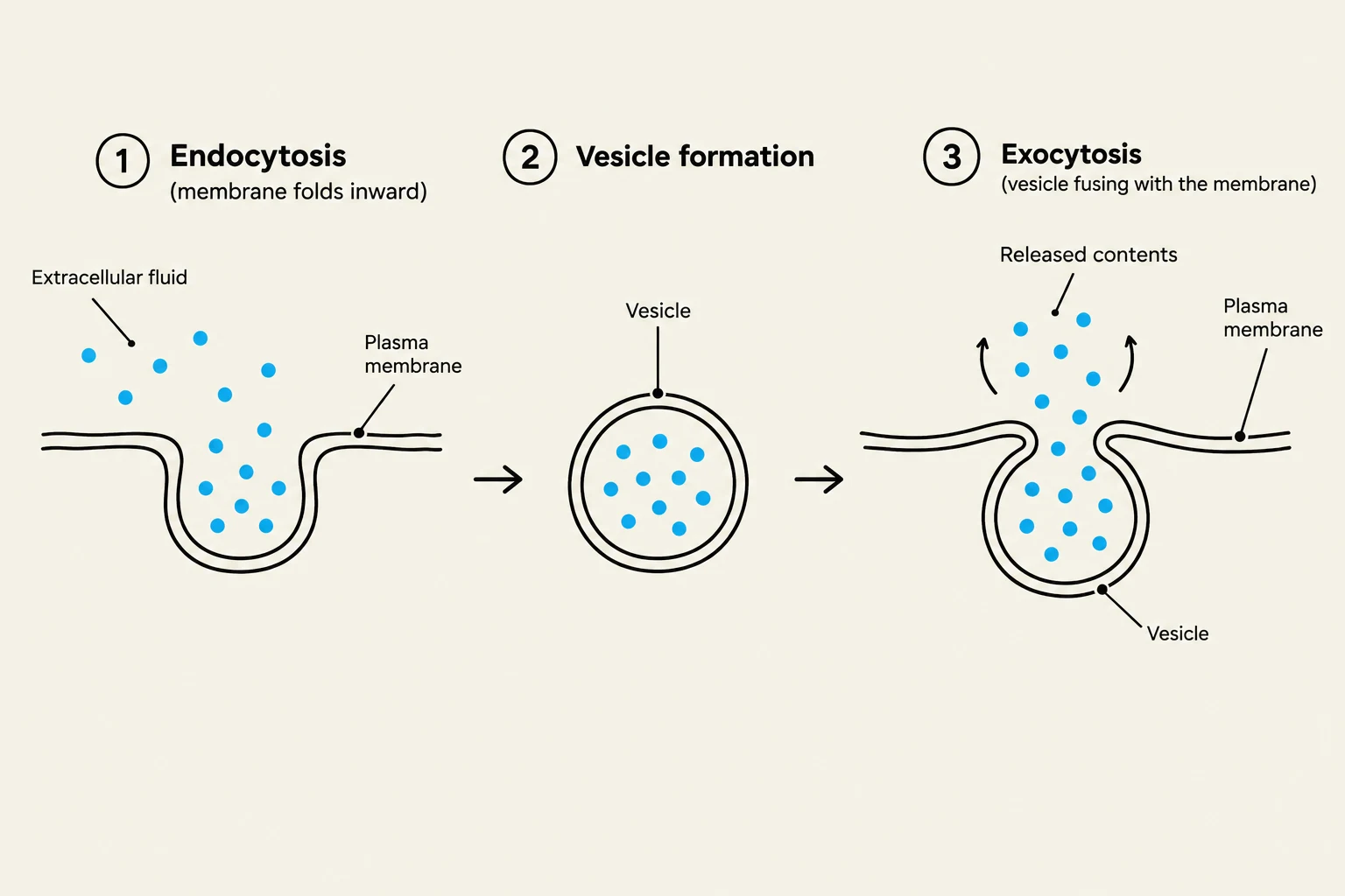
When the cell needs to move large quantities of material, it utilizes vesicular transport, which relies on the membrane's ability to undergo membrane fusion and self-healing.
Endocytosis: The membrane invaginates (folds inward) to capture external materials, pinching off to form a vesicle inside the cell.
Exocytosis: Vesicles (often from the Golgi apparatus) move toward the membrane, fuse with it, and release their contents to the outside.
Self-healing: Because of the hydrophobic effect, if a membrane is punctured, the hydrophobic tails will spontaneously move to avoid water, causing the edges to close and "heal" the gap.
Diagram illustrating the processes of endocytosis, vesicle formation, and exocytosis in a cell.
Cell-to-Cell Junctions
Cells in multicellular organisms do not exist in isolation; they are connected by specialized junctions that provide adhesion, communication, or sealing:
Junction Type
Primary Function
Key Protein/Structure
Tight Junctions
Create a waterproof seal between cells.
Claudins/Occludins
Adherens Junctions
Provide mechanical anchorage via the actin cytoskeleton.
Cadherins + Actin
Desmosomes
Provide high mechanical strength via intermediate filaments.
Cadherins + Intermediate Filaments
Gap Junctions
Allow direct chemical/electrical communication between cytoplasms.
Connexins (forming channels)
Clinical Connection: In cardiac muscle, gap junctions are essential. They allow calcium ions to flow directly from one cell to the next, creating a "domino effect" of electrical signals that ensures the entire heart contracts in a synchronized rhythm.
Microscopy: Visualizing the Membrane
Because cell membranes are incredibly thin, they cannot be seen with standard light microscopy. Scientists use advanced techniques to study them:
Transmission Electron Microscopy (TEM): Uses electron beams to see the internal structure and the bilayer layers.
Scanning Electron Microscopy (SEM): Provides detailed images of the cell surface.
Fluorescence Microscopy: Uses fluorescent markers attached to specific proteins to observe how molecules diffuse and move within the fluid membrane.
Keep reading for free.
Create an account to finish this reading and make it part of your study routine.