Cell Communication: Receptors and Signal Transduction
Explore the fundamental mechanisms of cell communication, including ligand-receptor interactions, signal transduction pathways, and the three primary classes of cell surface receptors (GPCRs, enzyme-linked receptors, and ligand-gated ion channels). Learn about second messengers, signal amplification, and different mode
In the complex environment of a multicellular organism, cells must constantly sense and respond to their surroundings to survive and function effectively. This ability to detect, interpret, and respond to external signals is known as cell communication. Whether it is a yeast cell increasing its metabolic enzymes in response to glucose or a plant bending toward light (phototropism), the fundamental mechanism remains the same: a signal is received, processed, and translated into a specific cellular action.
The Mechanics of Ligand Binding
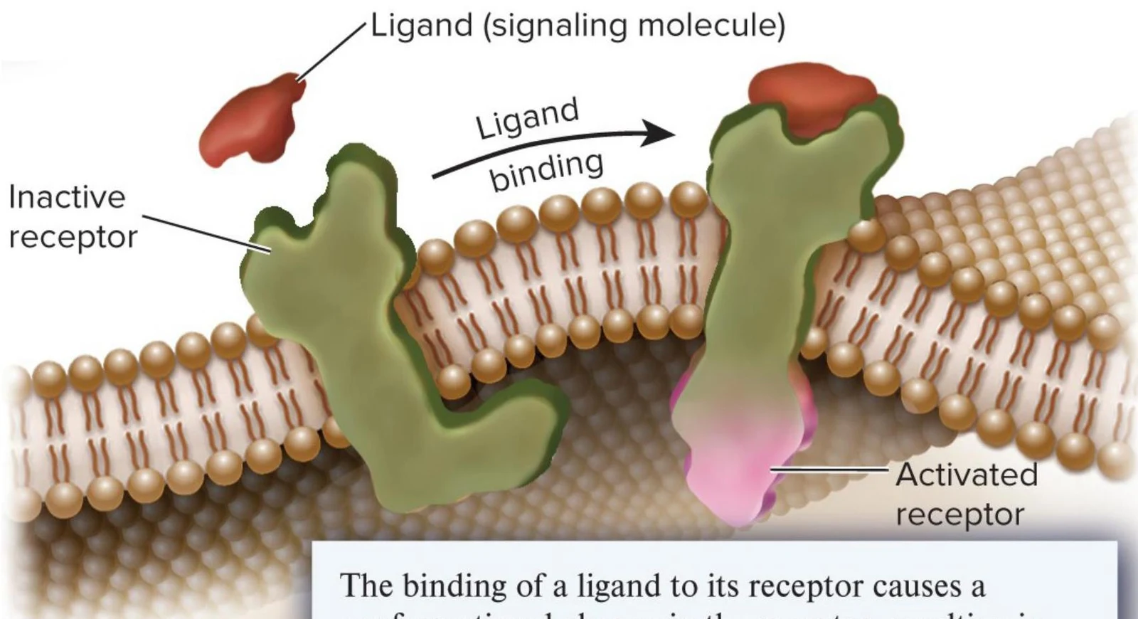
At the heart of cell communication is the interaction between a ligand (a signaling molecule) and a receptor (a specialized protein). Unlike enzymes, which perform catalysis by transforming a substrate into a different chemical product, receptors typically do not alter the chemical structure of their ligands. Instead, the ligand binds to the receptor, and the primary outcome is a conformational change—a shift in the receptor's 3D shape that activates its function.
The relationship between a ligand and its receptor is defined by high specificity and reversible binding. This is often explained through two models:
Lock-and-key model: The ligand fits perfectly into the receptor's binding site like a key into a lock.
Induced-fit model: The binding of the ligand induces the receptor to adjust its shape to fit the ligand more tightly.
The strength of this interaction is measured by the dissociation constant (Kd). The relationship between the concentrations of the ligand, the receptor, and the resulting complex is expressed as:
[Ligand⋅Receptor complex][Ligand][Receptor]=konkoff=Kd
Where kon is the rate of binding and koff is the rate of dissociation. A lower Kd indicates a higher affinity between the ligand and the receptor.
Illustration showing a ligand binding to an inactive receptor on a cell membrane, leading to an activated receptor.
Three Primary Classes of Cell Surface Receptors
Because many signaling molecules are too large or too hydrophilic to cross the plasma membrane, they must bind to receptors located on the cell surface. There are three main types of these receptors:
1. G-Protein-Coupled Receptors (GPCRs)
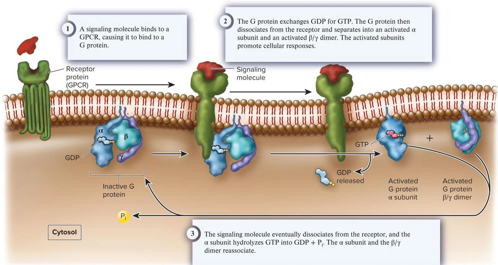
GPCRs are common in eukaryotes and are characterized by having seven transmembrane segments. When a ligand binds to a GPCR, the receptor activates a membrane-associated G protein. This activation involves the G protein exchanging GDP for GTP, causing the protein to dissociate into an α subunit and a β/γ dimer, which then trigger downstream responses.
This diagram illustrates the process of G protein-coupled receptor (GPCR) signaling, where a signaling molecule binds to a GPCR, activating a G protein that then initiates cellular responses.
2. Enzyme-Linked Receptors
These receptors possess an extracellular domain for ligand binding and an intracellular domain with catalytic function. A prominent example is the Receptor Tyrosine Kinase (RTK), such as the Epidermal Growth Factor (EGF) receptor. Upon ligand binding, these receptors often dimerize and activate their catalytic domain, which can then phosphorylate intracellular proteins.
Diagrams illustrating the structure and function of enzyme-linked receptors in cell signaling, showing how a signaling molecule binding to the receptor activates its intracellular catalytic domain to phosphorylate an intracellular protein using ATP.
3. Ligand-Gated Ion Channels
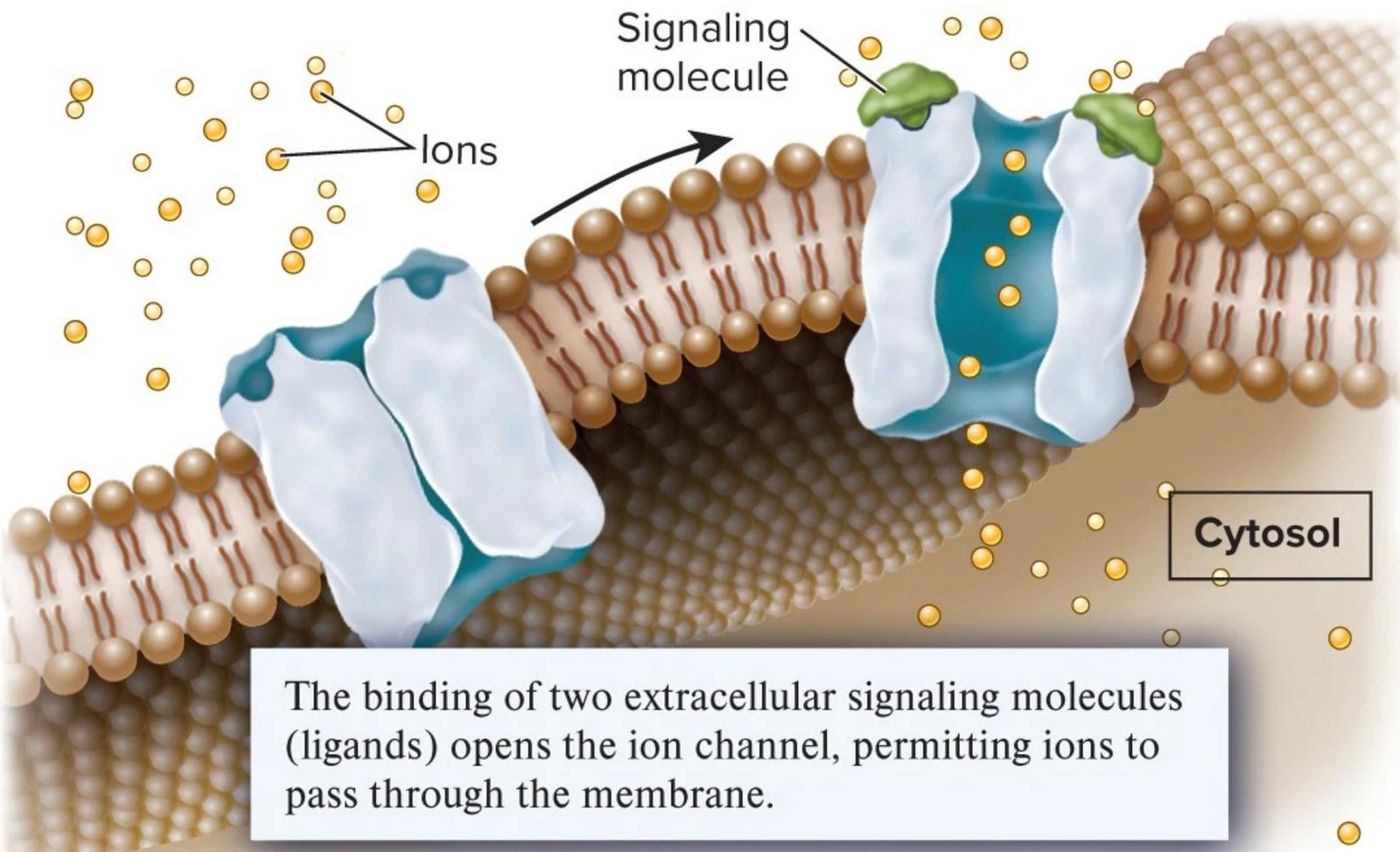
These receptors function as pores that open or close in response to the binding of a ligand. When the ligand binds, the channel changes shape, allowing specific ions to flow across the membrane. In animals, this mechanism is vital for transmitting signals between neurons and muscles.
This illustration depicts how signaling molecules bind to an ion channel in a cell membrane, causing it to open and allow ions to pass through into the cytosol.
Note on Intracellular Receptors: Not all receptors are on the surface. Steroid receptors are located in the cytosol or nucleus. Because steroid hormones are lipophilic, they can diffuse directly through the plasma membrane to bind these receptors, which then act as transcription factors to regulate gene expression.
Signal Transduction and Cellular Response
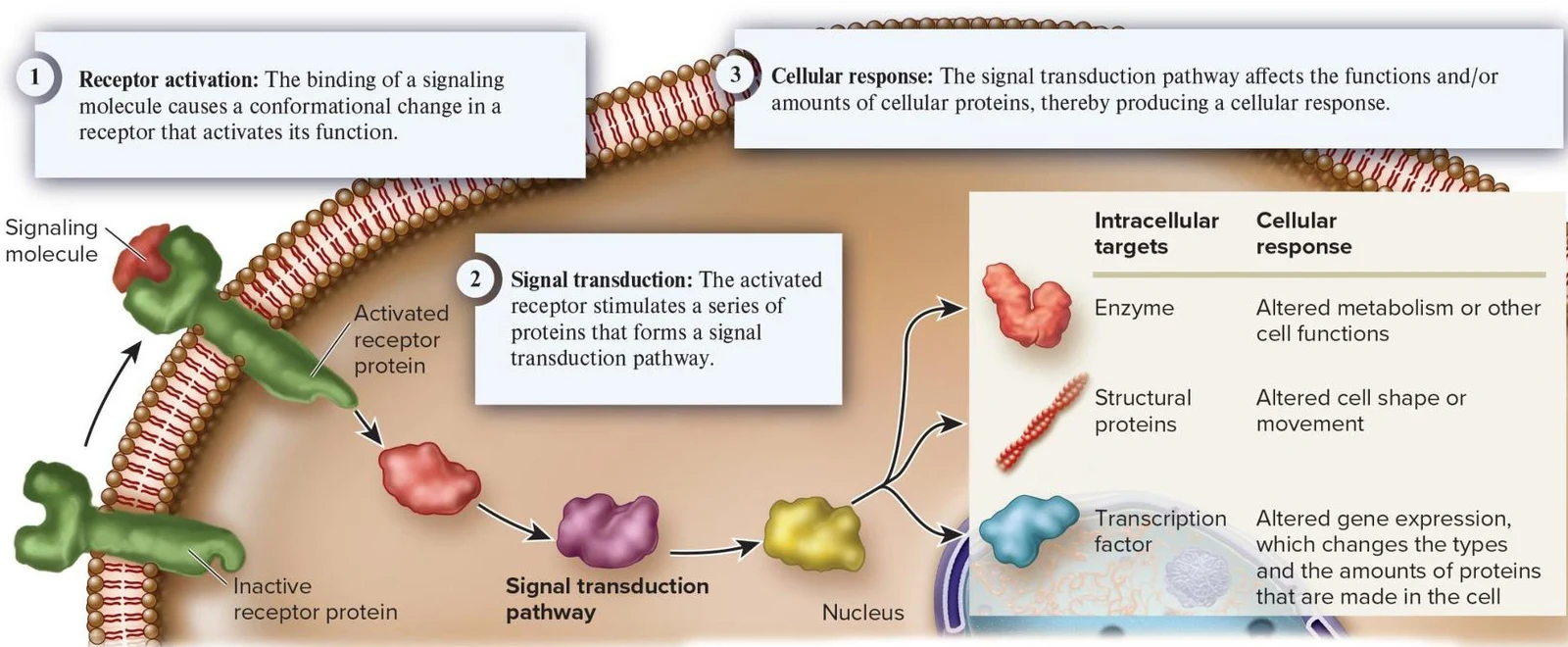
The process of converting an extracellular signal into a cellular response occurs in three distinct stages:
Receptor Activation: The ligand binds to the receptor, inducing a conformational change.
Signal Transduction: The activated receptor triggers a signal transduction pathway, a series of protein interactions that relay the message.
Cellular Response: The pathway targets specific components to produce an outcome.
This diagram illustrates the process of signal transduction, where a signaling molecule binds to a receptor, initiating a cascade of events that leads to a cellular response, such as altered metabolism, cell shape, or gene expression.
Second Messengers and Amplification
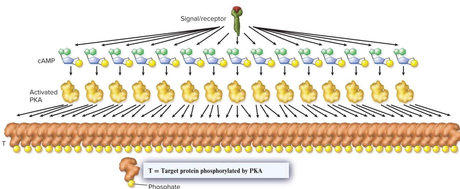
Many pathways utilize second messengers—small, non-protein, water-soluble molecules like cyclic AMP (cAMP) or calcium ions. Second messengers offer two major advantages: speed (they diffuse rapidly) and amplification (one receptor can trigger the production of many second messenger molecules, which in turn activate many target proteins).
This diagram illustrates the signal transduction pathway initiated by a signal/receptor, leading to the activation of cAMP and subsequently PKA. Activated PKA then phosphorylates target proteins.
The cellular response typically targets one of three types of proteins:
Structural Proteins: Altering cell shape or movement.
Transcription Factors: Altering gene expression to change the types and amounts of proteins in the cell.
Crosstalk
Cells rarely rely on a single isolated pathway. Instead, they utilize crosstalk, where components of one signal transduction pathway interact with—either activating or inhibiting—another pathway. This allows the cell to integrate multiple environmental inputs into a single, unified response.
Modes of Cellular Communication
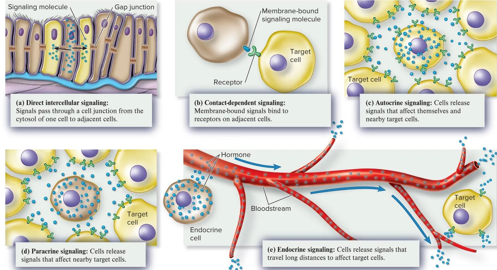
Cells communicate over varying distances using different mechanisms:
Signaling Type
Distance
Mechanism
Direct Intercellular
Immediate
Passing through cell junctions like gap junctions
Contact-Dependent
Adjacent
Membrane-bound ligands bind to receptors on a touching cell
Autocrine
Self
Cell releases signals that bind to its own surface
Paracrine
Local
Signals influence nearby target cells
Endocrine
Systemic
Hormones travel through the bloodstream to distant targets
This illustration depicts five types of cell signaling: direct intercellular signaling, contact-dependent signaling, autocrine signaling, paracrine signaling, and endocrine signaling.
Exam Tip: Remember that while enzymes transform substrates into products, receptors transform a signal into a conformational change. Both use high specificity and the lock-and-key/induced-fit models, but their "end products" are fundamentally different.
Keep reading for free.
Create an account to finish this reading and make it part of your study routine.