Cell Biology: Organelles, Protein Synthesis, and Degradation
An overview of cell biology, covering the structure and function of key organelles like the nucleus, endoplasmic reticulum, and Golgi apparatus. It explains protein synthesis, post-translational modifications, the secretory pathway, and mechanisms of protein degradation via proteasomes and lysosomes.
The cell is the fundamental unit of life, a complex and highly organized system enclosed by a lipid bilayer membrane. To function effectively, cells rely on specialized compartments known as organelles and various subcellular structures. The internal environment of the cell is divided into the cytosol (the fluid component) and the cytoplasm (the sum of the cytosol and all organelles). Understanding how these components interact—specifically how proteins are synthesized, modified, and eventually degraded—is central to the study of cell biology.
The Genetic Command Center: The Nucleus
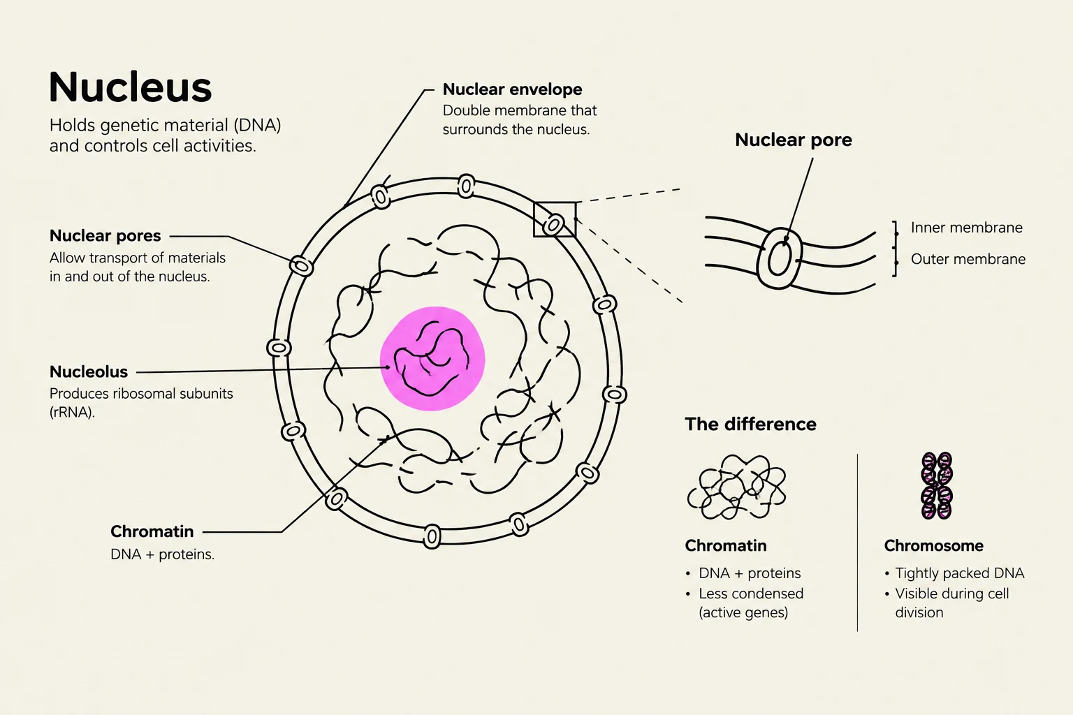
The nucleus serves as the repository of genetic information. It is enclosed by the nuclear envelope, which consists of two membranes that create a perinuclear cisternal space. This envelope is interrupted by nuclear pores, which mediate the translocation of molecules between the nucleus and the cytoplasm.
Inside the nucleus, DNA is organized into chromatin, a complex of DNA and proteins (including histones and non-histone proteins). Chromatin exists in two functional states:
Heterochromatin: Highly condensed and transcriptionally inactive.
Euchromatin: Loosely arranged and transcriptionally active.
A prominent non-membrane-bound structure within the nucleus is the nucleolus (plural: nucleoli). The nucleolus is the primary site for rRNA synthesis and the initial assembly of ribosomes.
Diagram illustrating the structure of a cell nucleus, including its components like the nuclear envelope, nuclear pores, nucleolus, and chromatin. It also shows the difference between chromatin and a chromosome.
Protein Synthesis and the Secretory Pathway
Proteins are the workhorses of the cell, and their destination determines which "pathway" they follow.
The Ribosome and the Rough ER
Translation begins on ribosomes, which are RNA-protein complexes consisting of small (40S) and large (60S) subunits. Ribosomes can be free in the cytosol or bound to the endoplasmic reticulum (ER).
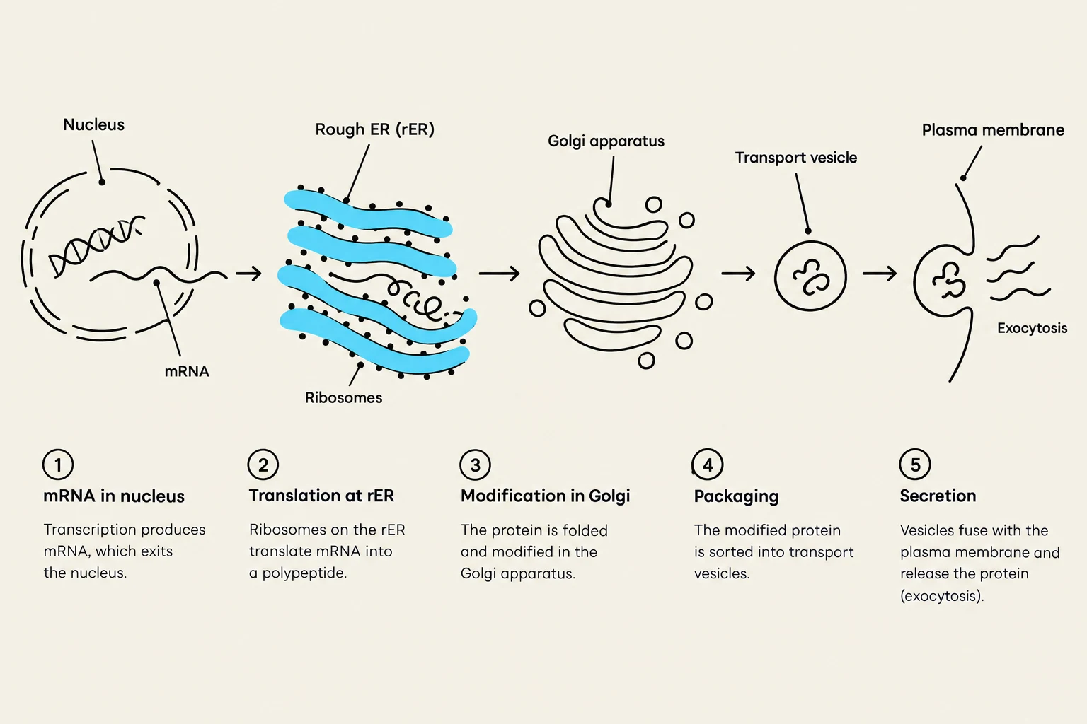
If a growing polypeptide contains a specific ER signal peptide, translation continues on the rough endoplasmic reticulum (rER). The rER is a system of interconnected sacs called cisternae, studded with ribosomes. This is the starting point for the secretory pathway, which handles proteins destined for secretion, the plasma membrane, or lysosomes.
Post-Translational Modifications in the ER
As proteins enter the rER, they undergo post-translational modifications—the covalent addition of functional groups to a polypeptide. A critical modification is N-linked glycosylation, where an oligosaccharide is added to the side chain of an Asparagine (Asn) residue. This process aids in protein folding and prevents aggregation. To ensure correct folding, chaperones (such as heat-shock proteins) bind to nascent polypeptides to assist them in reaching their functional shape.
The Golgi Apparatus: Sorting and Packaging
Once properly folded, proteins are transported via vesicles from the rER to the cis-Golgi network in an anterograde transport process. The Golgi apparatus consists of stacked, flattened cisternae. It serves as the cell's "post office," performing further post-translational modifications, such as completing N-linked glycosylation and initiating O-linked glycosylation.
O-linked glycosylation is vital for protein stability and recognition, such as in mucins or cell-surface glycoproteins used for adhesion and signaling. Finally, proteins bud from the trans-Golgi network in vesicles to be sent to their final destinations.
This diagram illustrates the process of protein synthesis and secretion in a cell, starting from transcription in the nucleus, translation on the rough ER, modification in the Golgi apparatus, packaging into vesicles, and finally secretion via exocytosis.
Clinical Correlation: Insulin Secretion
In pancreatic β-cells, insulin is synthesized via the secretory pathway. Preproinsulin is translated on the rER, where the signal peptide is cleaved to form proinsulin. After processing in the Golgi, it is released into the bloodstream via exocytosis.
The Non-Secretory Pathway and Other Organelles
Not all proteins are destined for secretion. The non-secretory pathway involves proteins translated by free cytosolic ribosomes that are subsequently targeted to the nucleus, mitochondria, peroxisomes, or remain in the cytosol.
Mitochondria and Peroxisomes
Mitochondria: Known as the "powerhouse" of the cell, they generate energy through oxidative phosphorylation. They are enclosed by two membranes: the Outer Mitochondrial Membrane (OMM) and the Inner Mitochondrial Membrane (IMM), which houses the electron transport chain. Mitochondria also contain their own circular DNA, RNA, and ribosomes.
Peroxisomes: These organelles are responsible for many oxidation reactions, particularly the catabolism of very-long-chain (VLC) fatty acids. They contain enzymes like catalase, which detoxifies hydrogen peroxide (H2O2) generated during metabolic processes.
Smooth Endoplasmic Reticulum (sER)
Unlike the rER, the smooth endoplasmic reticulum (sER) lacks ribosomes. It is specialized for lipid synthesis (including steroids and phospholipids), carbohydrate metabolism, and the detoxification of drugs via enzymes like Cytochrome P450. In muscle cells, a specialized form of sER called the sarcoplasmic reticulum sequesters Ca2+ ions for contraction and relaxation.
Protein Degradation: Quality Control
Cells must constantly remove damaged, misfolded, or unnecessary proteins to maintain homeostasis. There are two primary systems for degradation:
1. The Proteasome (Ubiquitin-Proteasome System)
The proteasome is a non-membrane-bound protein complex located predominantly in the cytosol. Proteins destined for destruction are first "tagged" with a small protein called ubiquitin (polyubiquitination). The proteasome then enzymatically degrades these tagged proteins.
2. The Lysosome
Lysosomes are membrane-bound organelles that perform enzymatic degradation of macromolecules. They use acid hydrolases to digest cellular structures (autophagy) or extracellular material brought in via endocytosis.
Exam Tip: Lysosomal Targeting
How does a lysosome know which enzymes to contain? Lysosomal enzymes are synthesized in the rER and must receive a specific mannose 6-phosphate (M6P) tag in the Golgi apparatus. This tag acts as a "destination label".
This diagram illustrates the cellular processes of protein degradation by the proteasome in the cytosol and the lysosome, highlighting the breakdown of polyubiquitinated proteins into peptides or amino acids for recycling or further processing.
Clinical Correlation: I-cell Disease
I-cell disease (Mucolipidosis type II) is a genetic disorder caused by a failure in the post-translational modification of lysosomal enzymes. Because the enzymes lack the M6P tag, they are not sent to the lysosome, leading to the accumulation of undigested "inclusion bodies" within the cell.
Keep reading for free.
Create an account to finish this reading and make it part of your study routine.