An overview of cataracts, including their manifestations, surgical interventions like phacoemulsification and IOL implantation, and essential preoperative and postoperative care to ensure successful outcomes and monitor for complications.
Cataracts represent one of the most common causes of vision impairment and blindness worldwide. As a progressive condition characterized by the clouding of the eye's natural lens, cataracts interfere with the clear passage of light to the retina, leading to significant functional limitations. Effective management requires a comprehensive approach that spans from early symptom recognition to precise surgical intervention and meticulous postoperative care.
Recognizing the Manifestations of Cataracts
A cataract is essentially an opacification of the crystalline lens. While the lens is normally clear to allow light to focus sharply on the retina, age-related changes or trauma can cause proteins within the lens to clump together, creating a "foggy" or "cloudy" appearance.
Patients typically present with a constellation of visual disturbances rather than sudden loss of sight. Key clinical manifestations include:
Gradual, painless blurring of vision: Unlike acute glaucoma, cataract progression is usually slow and does not cause pain.
Glare and Halos: Patients often report significant difficulty driving at night due to light scattering caused by the cloudy lens.
Decreased Color Perception: Colors may appear faded, yellowed, or brownish as the lens matures.
Second Sight: Occasionally, a patient may experience a temporary improvement in near vision (myopic shift) as the lens density changes, though this is a deceptive sign of progression.
Exam Tip: If a patient presents with sudden, painful vision loss, think of acute angle-closure glaucoma or retinal detachment rather than a cataract. Cataracts are characteristically painless.
Diagrams illustrating the difference between a normal eye and an eye with a cataract, showing how light is focused on the retina in each case.
Surgical Interventions
When visual impairment begins to interfere with a patient's activities of daily living (ADLs)—such as reading, driving, or recognizing faces—surgical intervention is indicated. Currently, surgery is the only definitive treatment for cataracts.
Phacoemulsification
The modern gold standard for cataract extraction is phacoemulsification. This technique involves several sophisticated steps:
Incision: A small incision is made in the cornea.
Fragmentation: An ultrasonic probe is inserted into the eye to vibrate at high frequencies, emulsifying (breaking up) the cloudy lens into tiny fragments.
Aspiration: The fragmented lens material is then suctioned out of the eye.
Intraocular Lens (IOL) Implantation
Once the natural lens is removed, the eye's refractive power is significantly altered. To restore vision, a synthetic Intraocular Lens (IOL) is implanted into the capsular bag. IOLs come in various forms, including:
Monofocal lenses: Provide clear vision at one distance (usually far).
Multifocal/Extended Depth of Focus (EDOF) lenses: Designed to provide vision at multiple distances, reducing the need for glasses.
Phacoemulsification is a modern cataract surgery technique that uses ultrasonic energy to break up the cloudy lens into tiny fragments, which are then aspirated, leaving a clear lens capsule.
Preoperative Care and Preparation
Successful surgical outcomes begin well before the patient enters the operating room. Preoperative management focuses on stabilizing the patient's ocular health and managing expectations.
Clinical Assessment:
Ocular Biometry: Precise measurements of the eye's axial length and corneal curvature are required to calculate the correct power of the IOL.
Medical Clearance: Given that many cataract patients are elderly, it is vital to assess cardiovascular stability and manage systemic conditions like diabetes, which can affect healing.
Patient Education:
Patients should be instructed on the "nothing in the eye" rule post-surgery and informed about the expected recovery timeline.
Postoperative Care and Complication Monitoring
Postoperative care is critical to prevent infection (endophthalmitis) and manage inflammation. The primary goal is to ensure the eye heals in a sterile, stable environment.
Nursing and Patient Interventions
Medication Adherence: Patients must strictly follow a regimen of topical antibiotic and corticosteroid eye drops to prevent infection and control inflammation.
Activity Restrictions: To prevent increased intraocular pressure (IOP) or displacement of the IOL, patients should avoid:
Bending at the waist (head below heart level).
Lifting heavy objects (>5-10 lbs).
Straining during bowel movements (Valsalva maneuver).
Protection: Wearing an eye shield at night is often required to prevent accidental rubbing or pressure on the eye during sleep.
Monitoring for Complications
While most surgeries are highly successful, clinicians must remain vigilant for signs of complications.
Complication
Clinical Manifestation
Endophthalmitis
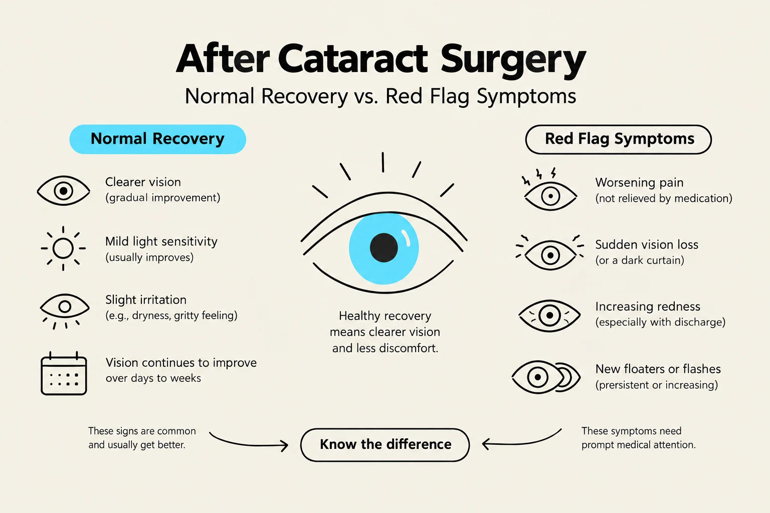
Severe pain, sudden redness, and rapid loss of vision (Medical Emergency).
Retinal Detachment
Sudden onset of floaters, flashes of light, or a "curtain" falling over the vision.
Ocular Hypertension
Increased pressure, often causing a dull ache or headache.
This infographic illustrates the normal recovery signs and red flag symptoms following cataract surgery, emphasizing the importance of knowing the difference for prompt medical attention.
Clinical Alert: If a patient reports a sudden increase in pain or a significant drop in vision following cataract surgery, they must be evaluated immediately to rule out endophthalmitis, which can lead to permanent blindness if not treated aggressively.
By integrating early recognition, advanced surgical techniques, and rigorous postoperative protocols, healthcare providers can significantly improve the quality of life for patients suffering from cataracts.
Keep reading for free.
Create an account to finish this reading and make it part of your study routine.