An educational overview of the vascular supply to the head and neck, detailing the anterior and posterior circulation pathways, the Circle of Willis, and the characteristics of the internal and external carotid arteries.
Understanding the vascular supply to the head and neck is fundamental to clinical practice, particularly in diagnosing stroke, carotid stenosis, and intracranial pathologies. The system is a complex arrangement of high-pressure arteries designed to deliver a constant, uninterrupted supply of oxygenated blood to the brain, the body's most metabolically demanding organ. This supply is achieved through two primary pathways: the anterior circulation (via the carotid system) and the posterior circulation (via the vertebral-basilar system).
The Aortic Arch and Carotid Origins
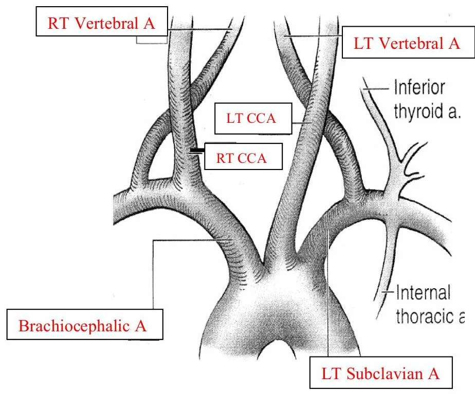
The journey of blood to the head begins at the aortic arch. The arch gives rise to three major branches that distribute blood to the upper body:
Brachiocephalic Artery: The first branch, which subsequently divides into the right subclavian and right common carotid arteries.
Left Common Carotid Artery (LT CCA): The second branch, supplying the left side of the head and neck.
Left Subclavian Artery (LT Subclavian): The third branch, supplying the left arm and contributing to the vertebral arteries.
The vertebral arteries themselves arise bilaterally from the subclavian arteries. As the Common Carotid Artery (CCA) ascends, it reaches a critical junction known as the carotid bulb. Located at the distal CCA and proximal Internal Carotid Artery (ICA), this area is the most common site for plaque formation due to the complex hemodynamics at the bifurcation.
An illustration of the major arteries branching from the aortic arch, including the brachiocephalic artery, left common carotid artery, right common carotid artery, left vertebral artery, right vertebral artery, inferior thyroid artery, internal thoracic artery, and left subclavian artery.
Internal vs. External Carotid Arteries
The CCA bifurcates into two distinct vessels: the Internal Carotid Artery (ICA) and the External Carotid Artery (ECA). These vessels are distinguished not just by their destination, but by their hemodynamic profiles.
The Internal Carotid Artery (ICA)
The ICA is the primary driver of the anterior circulation. It is characterized by low resistance blood flow, meaning the flow is continuous and steady throughout both systole and diastole. This pattern is essential to ensure the highly vascular brain tissue receives constant perfusion regardless of the cardiac cycle.
The ICA is divided into four anatomical sections:
Cervical (Extracranial): The longest portion, located in the neck; notably, it has no branches at this level.
Petrous: Located within the temporal bone.
Cavernous (Carotid Siphon): An S-shaped section within the cavernous sinus. This section gives rise to the ophthalmic artery, the first major branch of the ICA. The ophthalmic artery further branches into the lacrimal (central retinal), supraorbital, frontal, and nasal (angular) arteries to supply the eyes, forehead, and nose.
Supraclinoid: The distal-most section where the ICA terminates by branching into the Anterior Cerebral Artery (ACA) and the Middle Cerebral Artery (MCA).
The External Carotid Artery (ECA)
In contrast, the ECA exhibits high resistance flow, which is pulsatile (tri- or biphasic) with a significantly decreased diastolic component. This is because the ECA supplies peripheral vascular beds (like the scalp and face) that do not require the constant, steady flow necessary for the brain.
The ECA is smaller than the ICA and is located anterior and medial to it. It provides eight major branches to the face, neck, and scalp:
Superior thyroid artery
Ascending pharyngeal artery
Lingual artery
Facial artery
Occipital artery
Posterior auricular artery
Maxillary artery
Superficial temporal artery
Exam Tip: If you are asked to differentiate the ICA from the ECA during a scan, remember that the ICA is larger, located posterior-lateral, has low resistance, and has no branches in the cervical region. The ECA is smaller, anterior-medial, has high resistance, and possesses multiple branches.
Anterior and Posterior Cerebral Circulation
The brain's blood supply is organized into two main systems that eventually converge.
Anterior Circulation
This system is supplied by the ICA. It terminates in two major cerebral arteries:
Middle Cerebral Artery (MCA): The largest component, carrying approximately $70%-80%$ of the brain's blood.
Anterior Cerebral Artery (ACA): Carries approximately $20%-30%$ of the brain's blood.
Posterior Circulation
The posterior circulation is supplied by the vertebral arteries, which travel up the neck and join to form the basilar artery. The basilar artery then supplies the posterior structures of the cranial cavity and eventually feeds into the Posterior Cerebral Artery (PCA).
The Circle of Willis and Collateralization
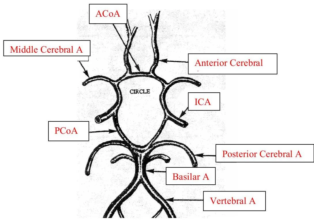
The convergence of the anterior and posterior pathways creates a sophisticated vascular network known as the Circle of Willis. This network is composed of the following vessels:
Anterior Cerebral Arteries (ACA)
Middle Cerebral Arteries (MCA)
Posterior Cerebral Arteries (PCA)
Basilar Artery
Anterior Communicating Artery (ACoA): Connects the left and right cerebral circulations.
Posterior Communicating Artery (PCoA): Connects the anterior circulation (ICA) to the posterior circulation (PCA).
Diagram of the Circle of Willis, illustrating the major arteries supplying blood to the brain.
The primary physiological importance of the Circle of Willis is collateralization. Collateralization is the ability of the vascular system to provide alternative pathways for blood flow. If one major vessel becomes stenosed (narrowed) or occluded (blocked), the communicating arteries allow blood to be rerouted from other vessels to ensure the brain remains perfused, thereby preventing ischemic damage.
Keep reading for free.
Create an account to finish this reading and make it part of your study routine.