Cardiovascular Physiology: Heart Function and Electrical Conduction
An overview of the cardiovascular system, detailing the dual-pump function of the right and left heart, hemodynamic principles, autonomic regulation, the heart's endocrine role, and the electrical conduction system.
The cardiovascular system is a sophisticated biological machine designed to transport oxygen, nutrients, and hormones to tissues while removing metabolic waste products. This process relies on a seamless integration of mechanical pumping, hemodynamic regulation, and precise electrical signaling. To understand how the heart maintains homeostasis, we must examine its anatomical divisions, the way it regulates blood pressure, its endocrine functions, and the specialized electrical cells that dictate the rhythm of life.
The Dual-Pump System: Right and Left Heart Function
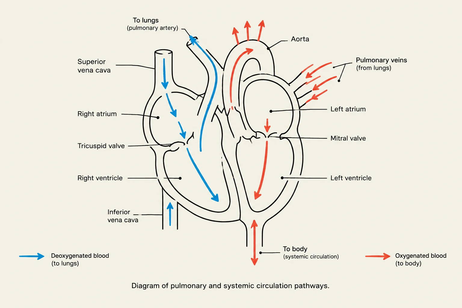
Diagram of pulmonary and systemic circulation pathways.
The heart functions as two separate pumps working in tandem to facilitate systemic and pulmonary circulation.
The Right Heart
The right side of the heart is responsible for the pulmonary circuit. It receives deoxygenated blood from the venous system and directs it toward the lungs. Blood flows through the tricuspid valve into the right ventricle, which then pumps the blood through the pulmonary valve into the pulmonary arteries for oxygenation.
The Left Heart
The left side of the heart manages the systemic circuit, which is significantly more demanding due to the resistance of the entire body. It receives oxygenated blood from the pulmonary system, which passes through the mitral (bicuspid) valve into the left ventricle. The left ventricle then pumps this oxygenated blood through the aortic valve into the aorta to deliver oxygen to the body. Because the left ventricle must pump against higher systemic resistance, it is a high-pressure chamber; excessive resistance (hypertension) can lead to left ventricular hypertrophy.
Hemodynamics and Autonomic Regulation
Cardiac performance is not static; it constantly adjusts to the metabolic demands of the body through the regulation of blood flow and pressure.
Measuring Cardiac Performance
Two fundamental equations govern our understanding of hemodynamics:
Cardiac Output (CO): This represents the volume of blood pumped by the heart per minute. It is the product of how much blood is pumped per beat and how fast the heart is beating.
CO=SV×HRWhere SV is Stroke Volume and HR is Heart Rate.
Blood Pressure (BP): This is the force exerted by blood against vessel walls, determined by the output of the heart and the resistance of the vessels.
BP=CO×SVRWhere SVR is Systemic Vascular Resistance.
Autonomic Modulation
The Autonomic Nervous System (ANS) acts as the "thermostat" for these values, utilizing different receptors to fine-tune cardiac and vascular activity:
Sympathetic Nervous System (Fight or Flight):
Alpha 1 (α1) receptors: Located in peripheral blood vessels; activation causes vasoconstriction, increasing SVR.
Beta 1 (β1) receptors: Located in the heart; activation increases both heart rate and the strength of contraction.
Beta 2 (β2) receptors: Located in the lungs and peripheral vessels; activation causes bronchodilation and vasodilation.
Parasympathetic Nervous System (Rest and Digest):
Mediated primarily by the vagus nerve (Cranial Nerve X), this system slows the heart rate and decreases atrioventricular conduction.
💡 Clinical Terminology Tip
When studying pharmacology or pathology, you will encounter these three terms regarding cardiac effects:
Chronotropy: Refers to Heart Rate (e.g., a positive chronotropic agent increases HR).
Inotropy: Refers to Contractile Strength (e.g., a positive inotropic agent increases contraction force).
Dromotropy: Refers to Conduction Speed (e.g., a negative dromotropic agent slows the electrical impulse).
The Heart as an Endocrine Organ
Beyond its mechanical role, the heart participates in hormonal regulation through the secretion of Natriuretic peptides. These hormones act to counter the Renin-Angiotensin-Aldosterone System (RAAS) to reduce blood volume and pressure:
Atrial Natriuretic Peptide (ANP): Released by atrial muscle cells in response to atrial distention (stretching). It reduces central venous pressure, cardiac output, and blood pressure.
Brain Natriuretic Peptide (BNP): Secreted primarily by the ventricles in response to excessive stretching of the myocytes.
C-Type Natriuretic Peptide (CNP): Secreted by the endothelium of blood vessels and serves a vasodilatory function.
Electrical Conduction and Action Potentials
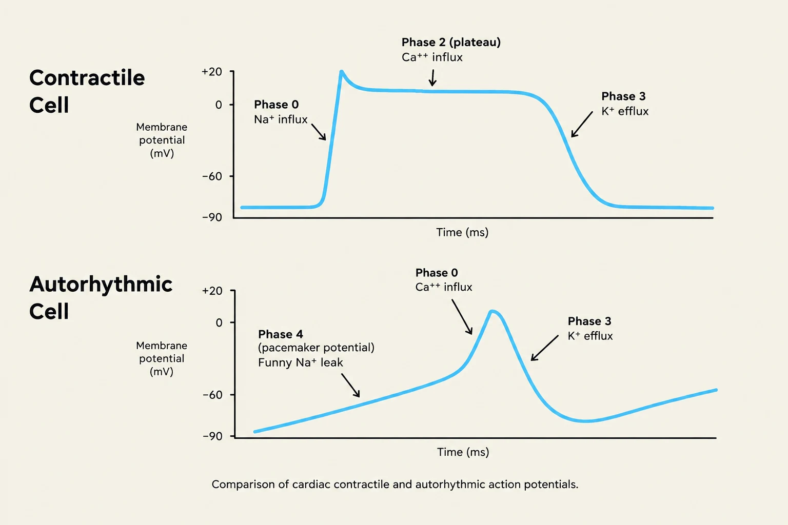
Comparison of cardiac contractile and autorhythmic action potentials.
The rhythmic contraction of the heart is driven by specialized cells that generate and conduct electrical impulses. These cells are categorized into two distinct types based on their function.
1. Cardiac Contractile Cells
These cells are responsible for the actual mechanical squeeze of the heart. Their action potential is characterized by specific ion movements:
Depolarization (Phase 0): Driven by a rapid influx of Sodium (Na+).
Plateau (Phase 2): A unique phase where Calcium (Ca++) enters the cell, facilitating the mechanical contraction.
Repolarization (Phase 3): Driven by the outward movement of Potassium ($K^+$), allowing the cell to relax.
2. Autorhythmic (Pacemaker) Cells
Unlike contractile cells, these cells do not require an external stimulus to fire; they generate their own rhythm:
Pacemaker Potential (Phase 4): A unique "funny" Na+ leak allows the membrane potential to drift upward toward the threshold.
Depolarization (Phase 0): Driven by the influx of Calcium (Ca++) rather than sodium.
Repolarization (Phase 3): Driven by the outward movement of Potassium ($K^+$).
The Hierarchy of Pacing
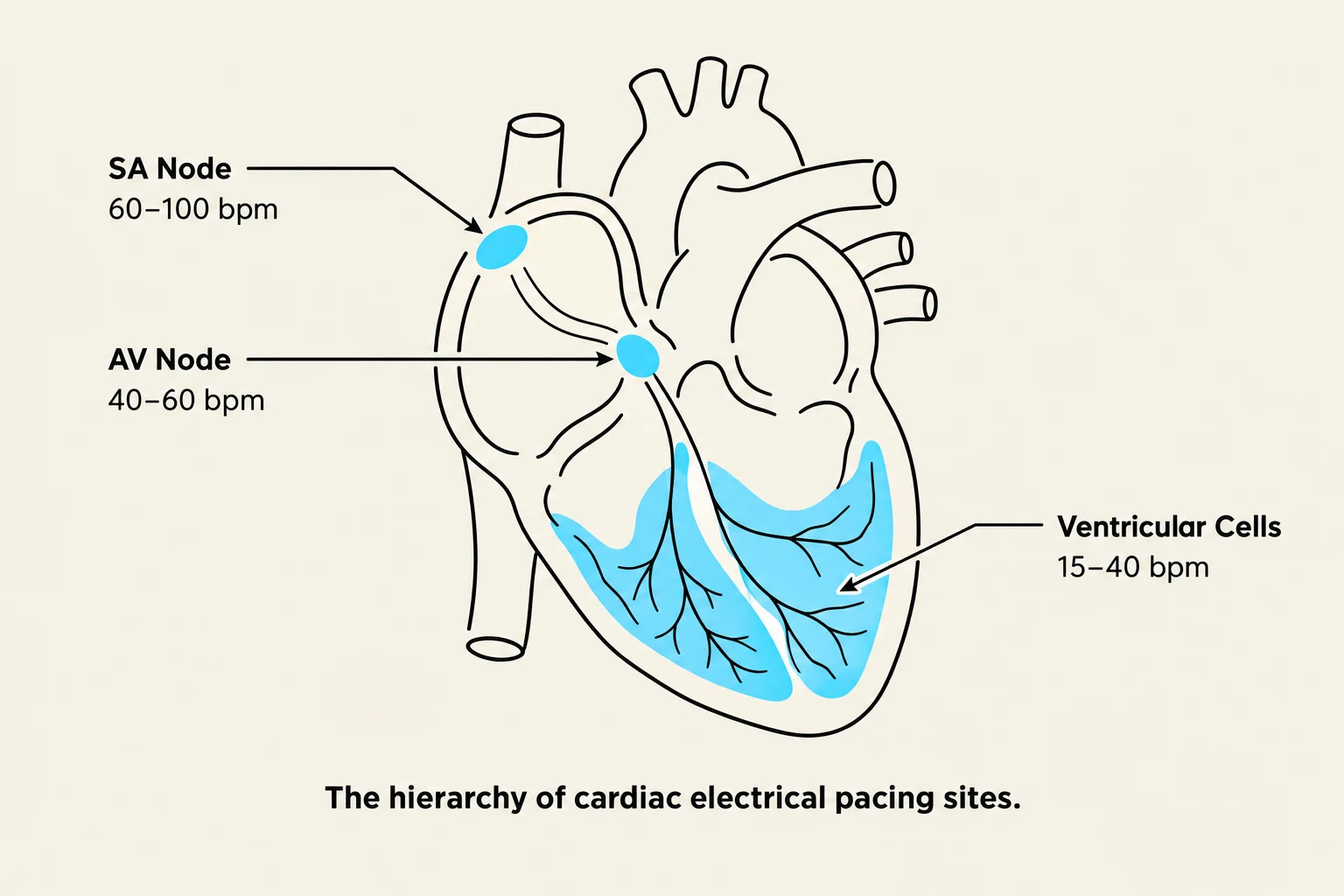
The hierarchy of cardiac electrical pacing sites, including the SA Node, AV Node, and Ventricular Cells, with their respective heart rate ranges.
The heart possesses a built-in hierarchy of electrical command. If one part of the system fails, the others can take over, though at a slower rate:
Pacing Site
Normal Heart Rate (bpm)
SA Node (Primary Pacemaker)
60 – 100
AV Node (Secondary Pacemaker)
40 – 60
Ventricular Cells (Tertiary Pacemaker)
15 – 40
⚠️ Exam Alert: Ion Differences
A common exam question involves distinguishing between the ions used in depolarization.
Contractile cells use Na+ for rapid depolarization.
Autorhythmic cells use Ca++ for depolarization.
Keep reading for free.
Create an account to finish this reading and make it part of your study routine.