Positron Emission Tomography (PET) has revolutionized nuclear cardiology by providing high-resolution imaging for the diagnosis of coronary artery disease (CAD), assessment of myocardial perfusion, and evaluation of myocardial viability. Unlike traditional SPECT imaging, PET utilizes radiopharmaceuticals that offer superior physiological insights through specific metabolic or molecular pathways. This article explores the production, physical properties, and biological mechanisms of three key PET tracers: N-13 Ammonia, F-18 Fluorodeoxyglucose (FDG), and F18 Flurpiridaz (Flycardo™).
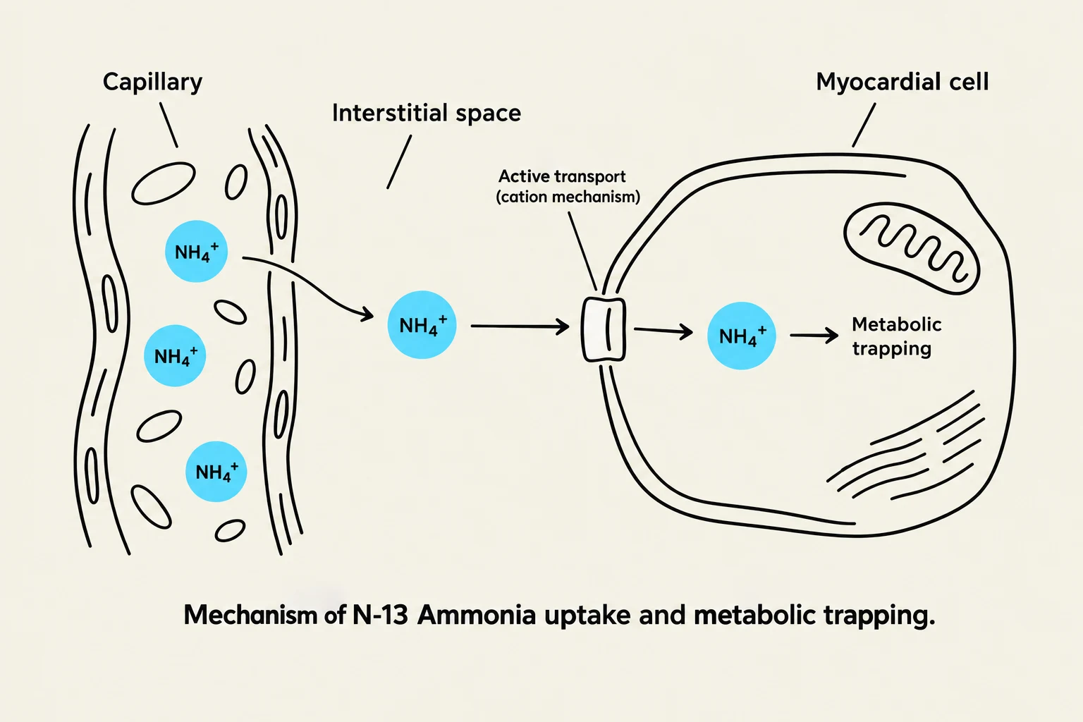
Diagram illustrating the mechanism of N-13 Ammonia uptake and metabolic trapping by a myocardial cell.
N-13 Ammonia: Perfusion and Rapid Kinetics
N-13 Ammonia is a primary agent used for myocardial perfusion imaging. Because of its short physical half-life, it requires on-site production via a cyclotron.
Production and Decay
The production of N-13 Ammonia involves a cyclotron reaction where a proton is absorbed by an Oxygen-16 target. The nuclear reaction can be summarized as follows:
16O+p+→17O−+4α→13N
. The resulting N-13 isotope decays via positron emission (β+) and has a very short physical half-life (T1/2p) of approximately 10 minutes.
Uptake Mechanism
N-13 Ammonia acts as an ammonium ion (NH4+). Its mechanism of entry into the myocardial cell is driven by an active transport cation mechanism. Once injected, the tracer rapidly exchanges across the capillary walls and transits through the interstitial spaces to reach the myocardial cells. A significant fraction of the tracer becomes "metabolically trapped" within the cells, allowing for stable imaging of perfusion. Due to its short half-life, redistribution is considered negligible.
Clinical Note: The critical organ for N-13 Ammonia is the bladder wall.
F-18 Fluorodeoxyglucose (FDG): Metabolic Imaging
While N-13 Ammonia tracks blood flow (perfusion), F-18 FDG is utilized to assess myocardial metabolism. This makes it an essential tool for distinguishing between viable tissue and scarred (infarcted) tissue.
Production and Physical Properties
F-18 FDG is a hydrophilic fluorinated sugar. It is produced in a cyclotron through the following reaction:
18O+p+→18F−β+→18O
. It possesses a significantly longer physical half-life (T1/2p) of 110 minutes compared to N-13 Ammonia, allowing for more flexible imaging protocols.
Mechanism of Action
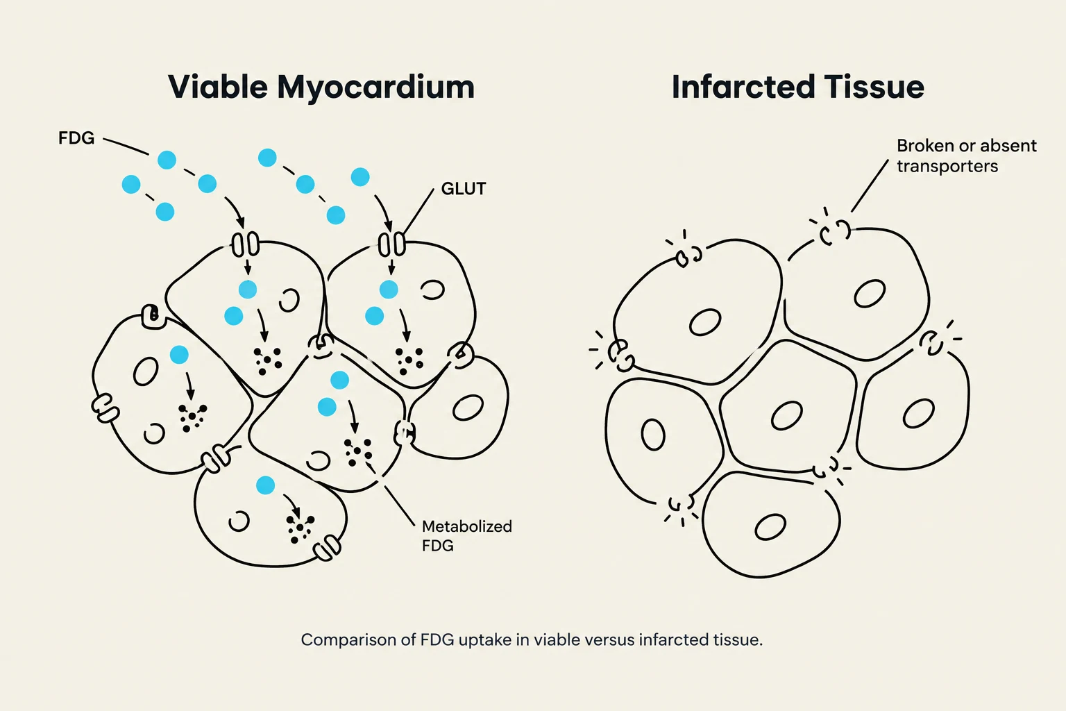
This diagram illustrates the difference in FDG uptake between viable and infarcted myocardial tissue. In viable myocardium, FDG is transported into cells via GLUT transporters and metabolized. In infarcted tissue, transporters are broken or absent, preventing FDG uptake.
The uptake of FDG is driven by cellular metabolism. As a glucose analog, the cells take up the tracer via glucose transporters. Once inside the cell, the tracer undergoes metabolic processes that allow it to be visualized during PET scanning. This metabolic activity is a hallmark of viable myocardium; cells that are alive continue to consume glucose, whereas dead or scarred tissue will show little to no FDG uptake.
F18 Flurpiridaz is a newer PET radiopharmaceutical designed to evaluate myocardial ischemia (MI) in patients with suspected coronary artery disease. It aims to combine the benefits of high extraction rates with the logistical advantages of longer half-lives.
Synthesis and Physical Properties
F18 Flurpiridaz has a physical half-life (T1/2p) of 110 minutes. Its synthesis is a complex two-step process:
Initial Reaction: Involves tetra-n-butylammonium fluoride (TBAF) in THF, tosyl chloride in 4-demethylaminopyridine (DMAP), triethylamine (TEA), and dichloromethane.
Fluorination: Treatment with potassium fluoride-18, kryptofix, and acetonitrile is used to increase the solubility of the fluoride ion in organic solvents.
Mitochondrial Mechanism and Viability
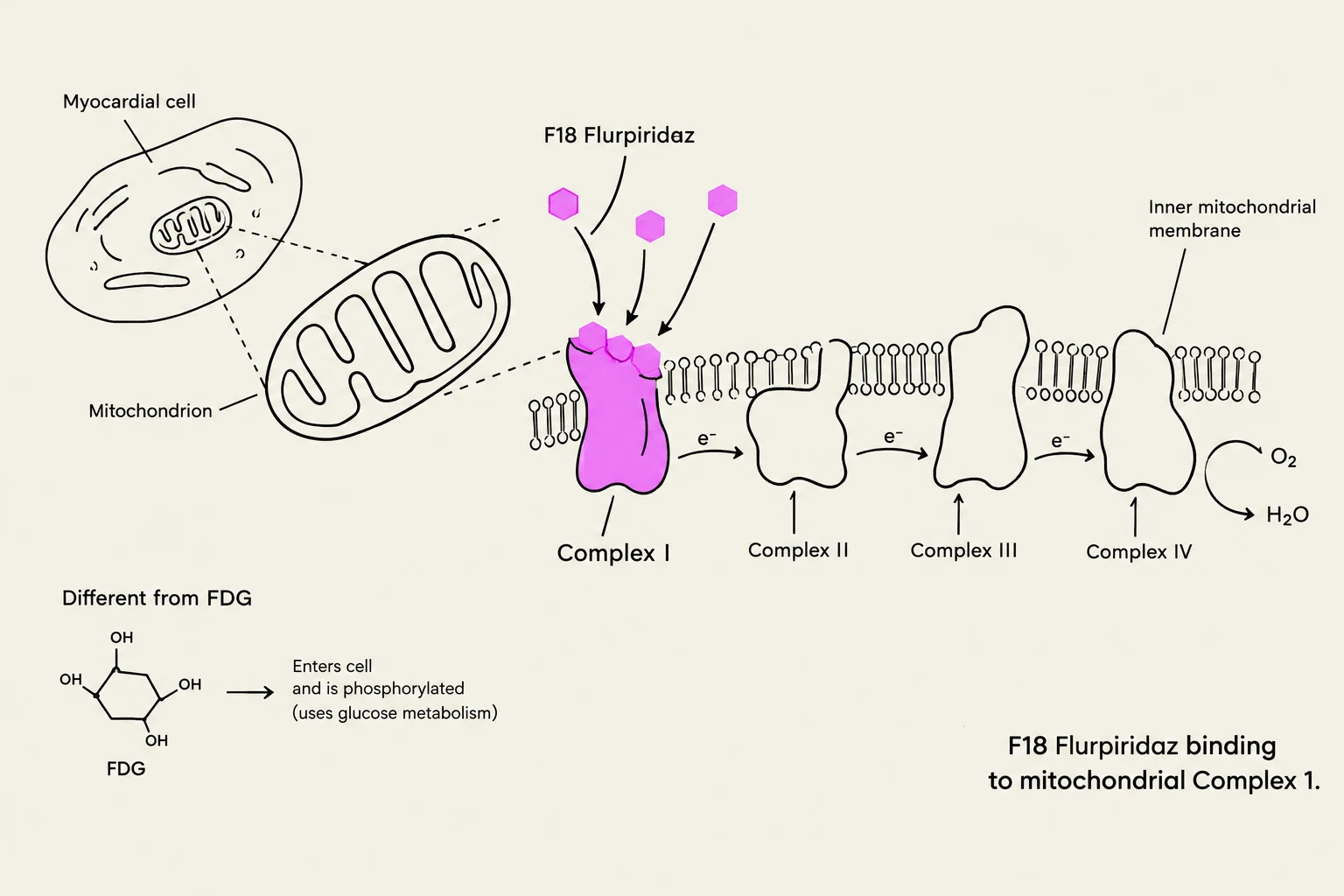
Diagram illustrating F18 Flurpiridaz binding to mitochondrial Complex 1, showing its interaction with the inner mitochondrial membrane and electron transport chain. Also depicts a myocardial cell and mitochondrion, and contrasts FDG's structure and cellular uptake via glucose metabolism.
The mechanism of F18 Flurpiridaz is unique. It serves as an analog of the mitochondrial complex 1 (MC-1) inhibitor, pyridaben. The tracer is extracted by the myocardium in direct proportion to myocardial blood flow and subsequently binds to heart tissue containing biologically active mitochondria.
This binding mechanism provides a clear distinction between tissue types:
Viable Myocardium: Contains active mitochondria, leading to high radioactivity uptake.
Infarcted Tissue: Lacks active mitochondria, resulting in low radioactivity.
Compared to other myocardial perfusion imaging (MPI) tracers, F18 Flurpiridaz offers a higher extraction rate and a lower effective dose to the patient.
Safety Protocol: The critical organ for F18 Flurpiridaz is the kidneys. The radiopharmaceutical must be discarded 8 hours after the end of synthesis or when the activity falls below the required injection level, whichever occurs first.
Summary Comparison of PET Tracers
The following table summarizes the key differences between the three discussed radiopharmaceuticals:
Property
N-13 Ammonia
F-18 FDG
F18 Flurpiridaz
Primary Use
Myocardial Perfusion
Myocardial Metabolism
Myocardial Perfusion/Ischemia
Mechanism
Active transport (cation)
Cellular metabolism
Mitochondrial Complex 1 binding
Half-life (T1/2p)
~10 minutes
110 minutes
110 minutes
Critical Organ
Bladder wall
N/A
Kidneys
Exam Tip: When asked to differentiate between perfusion and viability, remember that N-13 Ammonia and Flurpiridaz primarily track blood flow/mitochondrial activity (perfusion), while FDG tracks the actual consumption of sugar (metabolism/viability).
Keep reading for free.
Create an account to finish this reading and make it part of your study routine.