The cardiovascular system is a complex network designed to maintain perfusion to vital organs. When the integrity of blood vessels, heart valves, or the surrounding tissues is compromised, the resulting pathophysiology can lead to systemic failure, localized ischemia, or life-threatening emergencies. This article explores the mechanisms behind peripheral vascular diseases, valvular dysfunctions, and inflammatory conditions of the heart.
Peripheral Vascular Diseases
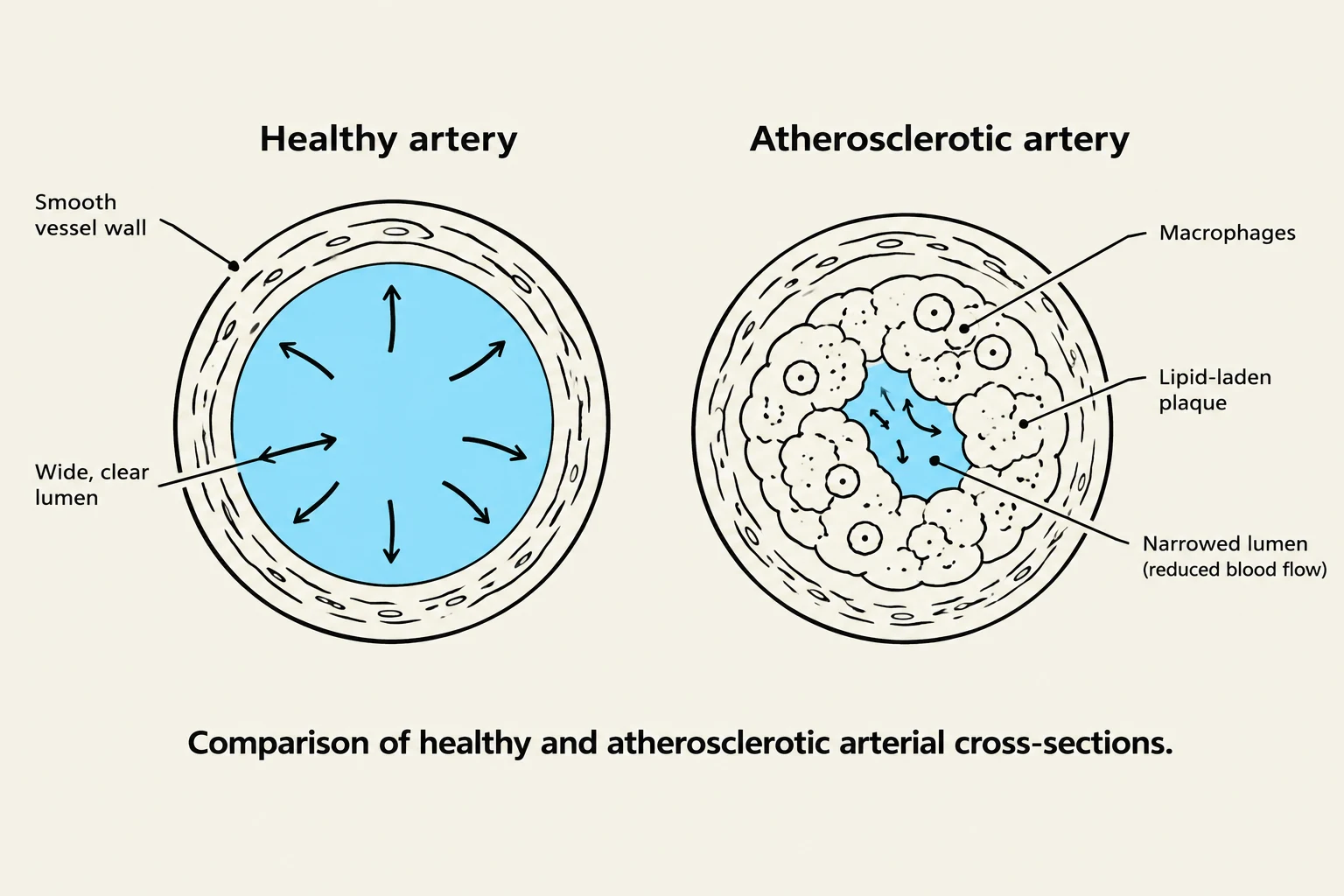
Comparison of healthy and atherosclerotic arterial cross-sections.
Peripheral vascular diseases involve alterations in the blood vessels that supply the limbs and other extremities. These conditions often stem from atherosclerosis—the thickening and hardening of arterial walls due to the accumulation of lipid-laden macrophages.
Thromboangiitis Obliterans (Buerger Disease)
Buerger disease is an inflammatory condition that affects small- and medium-sized arteries. It is most commonly observed in young male smokers. The disease is characterized by the formation of thrombi and intense vasospasm within the affected segments, which can lead to pain, tenderness, hair loss, and eventually gangrenous lesions.
Raynaud Phenomenon and Disease
Raynaud's involves episodic vasospasm in the digital arteries (fingers and toes), leading to ischemia. This manifests as distinct color changes: pallor, cyanosis, and redness as blood returns.
Raynaud Phenomenon: This is secondary to other systemic diseases or conditions.
Raynaud Disease: This is a primary vasospastic disorder of unknown origin, which tends to affect young women and is often triggered by cold exposure or emotional stress.
Peripheral Artery Disease (PAD)
PAD is primarily caused by the atherosclerotic obstruction of arteries that perfuse the limbs, particularly the lower extremities. A hallmark clinical manifestation is intermittent claudication, which is pain that occurs during ambulation due to inadequate blood flow. Severe cases may present with the "6 Ps": pallor, pulselessness, pain, paresthesias, paralysis, and poikilothermia.
Valvular Disorders
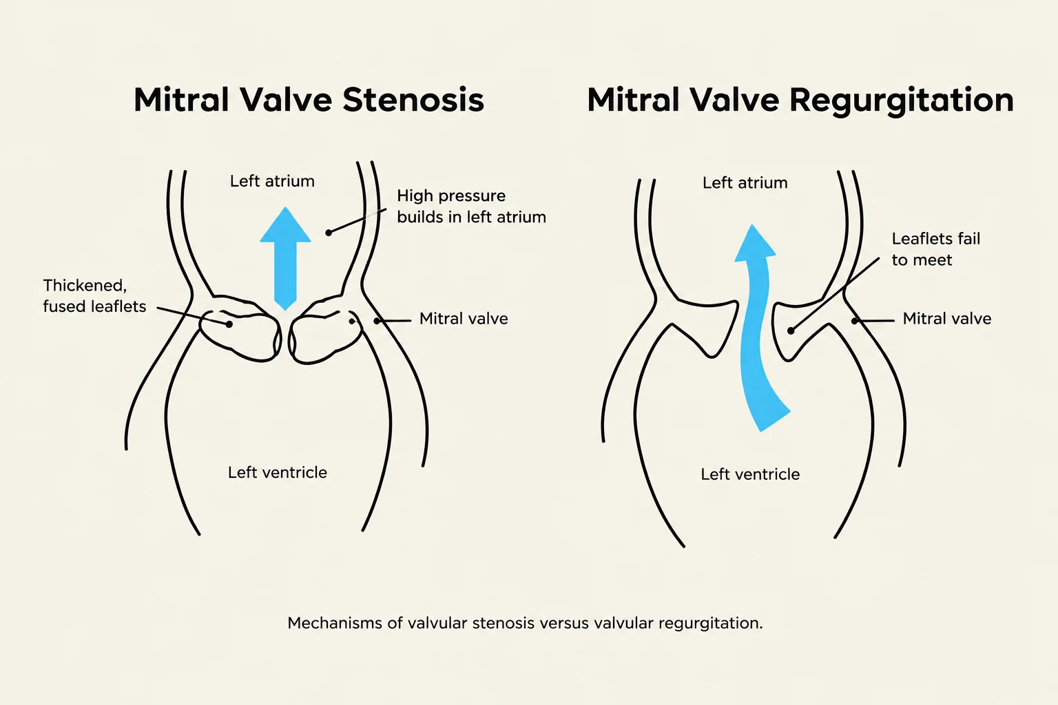
Diagrams illustrating the mechanisms of mitral valve stenosis and mitral valve regurgitation.
Heart valves ensure unidirectional blood flow. When valves fail, the heart must compensate for either an obstruction to flow (stenosis) or a failure to prevent backflow (regurgitation). These dysfunctions lead to increased workload, which can cause chamber dilation and myocardial hypertrophy.
Valvular Stenosis
Stenosis occurs when the valve orifice is narrowed, impeding forward blood flow and increasing pressure in the preceding chamber.
Aortic Stenosis: The most common valvular abnormality. It causes diminished blood flow from the left ventricle to the aorta, often resulting in angina, syncope, and a crescendo-decrescendo systolic murmur.
Mitral Stenosis: This impairs flow from the left atrium to the left ventricle, frequently caused by rheumatic fever. It is characterized by an "opening snap" and a diastolic murmur.
Pulmonic Stenosis: Often a result of congenital anomalies, presenting with a systolic murmur.
Valvular Regurgitation (Insufficiency)
Regurgitation occurs when the valve leaflets fail to close completely, allowing blood to flow backward.
Aortic Regurgitation: Results in a widened pulse pressure and a diastolic murmur.
Mitral Regurgitation: Permits backflow from the left ventricle to the left atrium. It is often caused by mitral valve prolapse or rheumatic heart disease and presents with a pansystolic murmur.
Tricuspid Regurgitation: Causes volume overload in the right heart and increased systemic venous pressure.
Exam Tip: When distinguishing between stenosis and regurgitation, remember that stenosis is an obstruction to forward flow (pressure builds behind the valve), while regurgitation is a failure of closure (volume increases behind the valve due to backflow).
Mitral Valve Prolapse (MVP) Syndrome
In MVP, the anterior and posterior cusps of the mitral valve billow upward into the atrium during systole. While often asymptomatic, patients with MVP are at a higher risk of developing infective endocarditis.
Inflammatory and Infectious Conditions
Acute Rheumatic Fever
This is an abnormal immune response to M proteins (often following a streptococcal infection) that cross-reacts with normal tissues. It can lead to rheumatic heart disease, characterized by permanent valvular damage. Key clinical manifestations include:
Aschoff bodies: Fibrinoid necrotic deposits found in the heart tissue.
Carditis: Inflammation of the heart.
Polyarthritis: Inflammation of large joints.
Chorea: Sudden, involuntary, irregular movements.
Infective Endocarditis
Diagram illustrating bacterial vegetations on a heart valve and their potential to enter the bloodstream.
Infective endocarditis is the inflammation of the endocardium caused by bacteria (such as Staphylococci or Streptococci) adhering to damaged valves. This adherence leads to the formation of vegetations—clumps of bacteria, fibrin, and platelets.
Clinical signs include fever, new or changed murmurs, and specific skin lesions:
Osler nodes: Painful, erythematous nodules on the pads of fingers and toes.
Janeway lesions: Nonpainful, hemorrhagic lesions on the palms and soles.
Pericarditis
Pericarditis is the inflammation of the pericardium. It typically presents as pleuritic chest pain that is worsened by inspiration or lying down, but improves when the patient leans forward.
Dressler’s Syndrome: A delayed form of post-myocardial infarction (MI) pericarditis that may occur weeks to months after the initial event, thought to be an antigen-antibody response to necrotic myocardium.
Summary Table of Valvular Murmurs
Valve
Stenosis Murmur
Regurgitation Murmur
Aortic
Crescendo-decrescendo systolic
Diastolic
Mitral
Diastolic (with opening snap)
Pansystolic
Tricuspid
Diastolic
Systolic (increases with inspiration)
Keep reading for free.
Create an account to finish this reading and make it part of your study routine.