Understanding the cardiovascular system requires a dual focus on the mechanical physics of blood movement (hemodynamics) and the biological processes that maintain or disrupt that movement. This article explores the fundamental principles of cardiac function, the mechanisms of vascular resistance, and the progression of atherosclerosis to provide a foundation for pharmacological intervention.
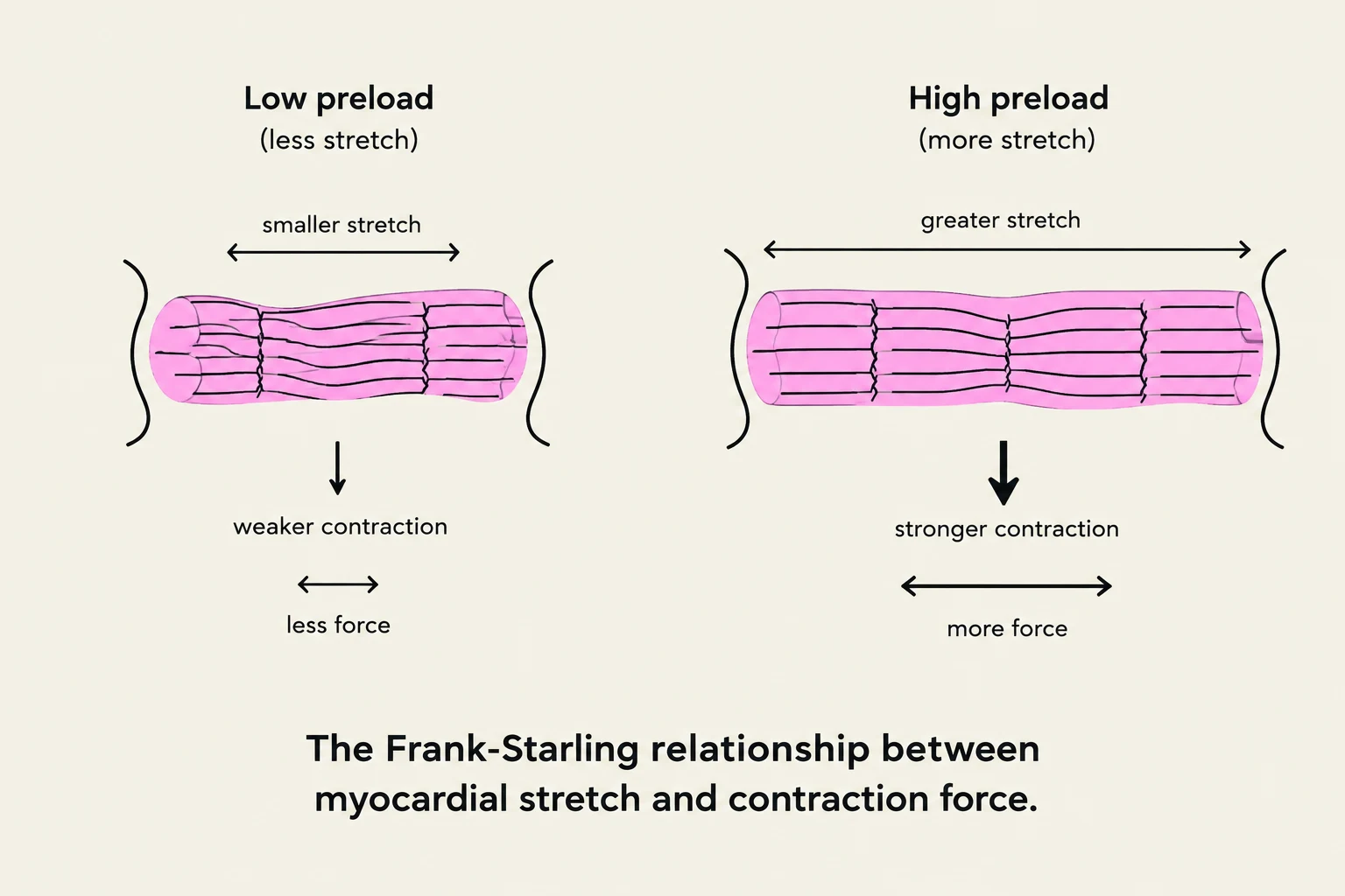
The Frank-Starling relationship illustrates how myocardial stretch affects contraction force, with greater stretch leading to stronger contractions.
Cardiovascular Physiology and Hemodynamics
The heart functions as a dual-pump system, moving blood through the pulmonary and systemic circuits. Blood flows through the right heart to the lungs for oxygenation, returns to the left heart, and is then pumped to the rest of the body. This flow is regulated by valves: atrioventricular (AV) valves manage flow between atria and ventricles, while semilunar valves (aortic and pulmonary) ensure unidirectional flow during ventricular contraction.
Cardiac Output and Stroke Volume
The efficiency of the heart is measured by Cardiac Output (CO), which is the total volume of blood pumped by the heart per minute. It is determined by the relationship between heart rate (HR) and stroke volume (SV):
CO=HR×SV
Stroke volume is the volume of blood ejected from the left ventricle during a single contraction. Several physiological determinants influence this volume:
Preload: This refers to the end-diastolic volume, or the degree of stretch on the ventricular muscle fibers at the end of filling.
Afterload: This is the resistance or pressure that the ventricle must overcome to eject blood into the systemic circulation.
The Frank-Starling Relationship: This principle states that as preload increases, the heart muscle stretches, leading to a more forceful contraction and increased stroke volume. However, this relationship only works "up to a point"; excessive stretching can eventually lead to decreased contractile efficiency.
Electrical Conduction and Autonomic Control
The rhythm of the heart is governed by specialized electrical tissues. The SA node acts as the natural pacemaker, initiating the impulse that travels through the conduction system. The AV node serves as a critical gatekeeper, delaying the impulse to allow the ventricles to fill before contraction.
The Autonomic Nervous System (ANS) modulates this electrical activity to meet the body's metabolic demands:
Sympathetic Nervous System: Releases neurotransmitters that increase both heart rate and myocardial contractility.
Parasympathetic Nervous System: Primarily acts via the vagus nerve to decrease heart rate and slow AV conduction.
Clinical Correlation: A medication that decreases heart rate (like a beta-blocker) or reduces circulating volume will typically decrease cardiac output.
Vessels, Resistance, and Blood Pressure
Blood pressure (BP) is not merely a single number but a product of the heart's output and the resistance encountered in the vessels:
BP=CO×SVR
(Where SVR is Systemic Vascular Resistance)
The Role of Arterioles and Resistance
Arterioles are the primary determinants of systemic vascular resistance. Because they have thick muscular walls, they can constrict or dilate to control blood flow. When the diameter of an arteriole decreases (vasoconstriction), resistance increases significantly, which in turn raises blood pressure.
Regulatory Systems
The body employs several mechanisms to maintain blood pressure homeostasis:
ANS and Baroreceptors: Rapid-response sensors that detect pressure changes and trigger immediate adjustments in heart rate and vessel diameter.
RAAS (Renin-Angiotensin-Aldosterone System): A slower, hormonal mechanism. Angiotensin II acts as a potent vasoconstrictor, while aldosterone promotes sodium and water retention by the kidneys, increasing blood volume and pressure.
Kidneys: Play a long-term role in regulating blood pressure through the management of fluid and electrolyte balance.
Atherosclerosis: From Injury to Plaque
While arteriosclerosis refers to a general hardening of the arteries, atherosclerosis is a specific type of arteriosclerosis involving the buildup of fatty plaques within the arterial walls.
The Pathophysiologic Cascade
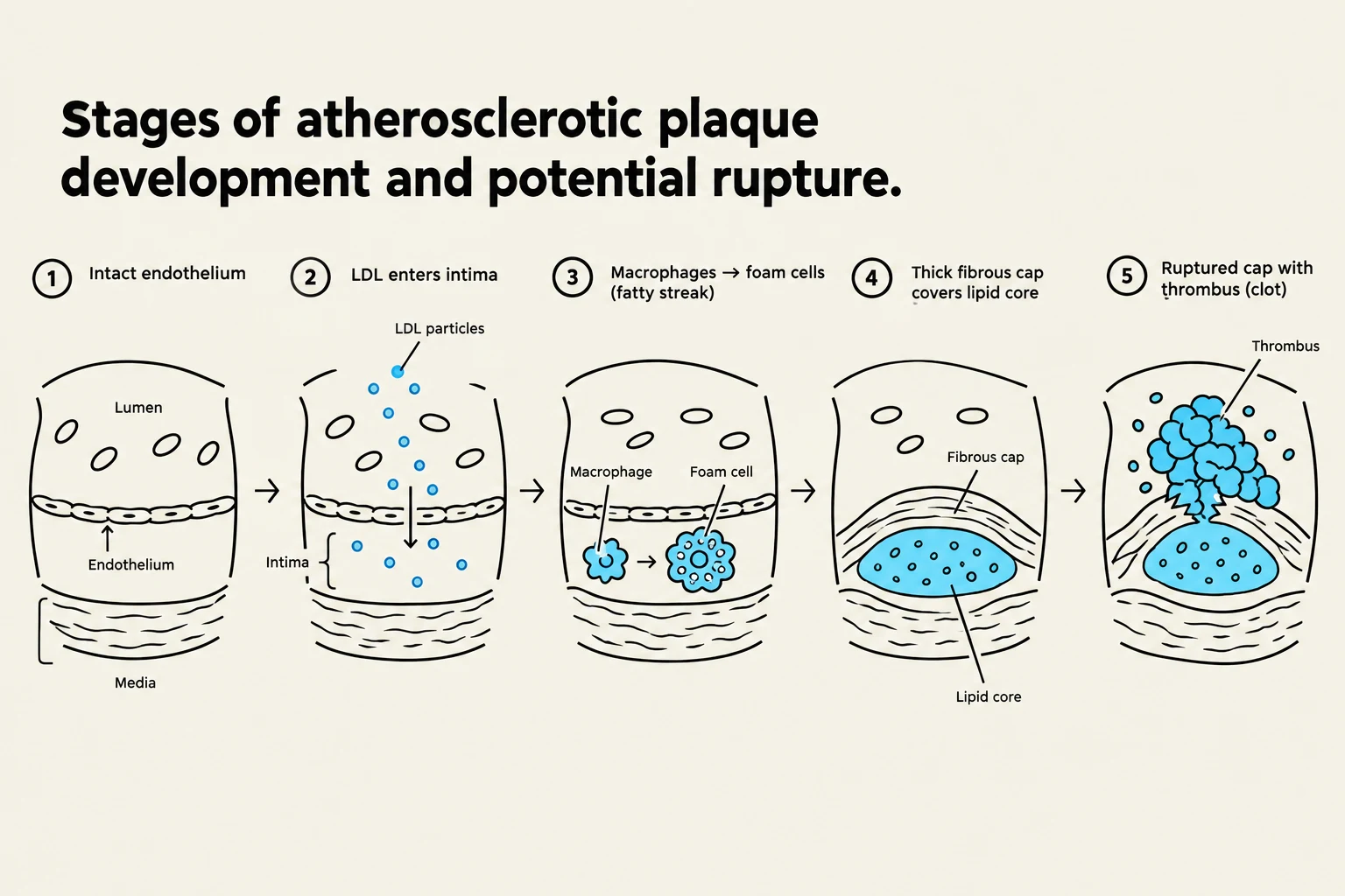
Diagram illustrating the five stages of atherosclerotic plaque development and potential rupture, from intact endothelium to a ruptured cap with thrombus.
The development of a plaque is a multi-stage inflammatory process:
Endothelial Injury: Triggered by risk factors such as hypertension, smoking, or hyperlipidemia.
Inflammation: The damaged endothelium becomes dysfunctional, allowing substances to enter the vessel wall.
LDL Oxidation: Low-density lipoproteins (LDL) enter the intima and become oxidized.
Foam Cell Formation: Macrophages engulf the oxidized LDL, transforming into "foam cells," which create a "fatty streak".
Fibrous Plaque: Smooth muscle cells proliferate and deposit collagen, creating a stable fibrous cap over the lipid core.
Plaque Rupture: If the cap becomes unstable and ruptures, it exposes thrombogenic material to the blood, triggering platelet activation and the formation of a clot (thrombus).
The clinical consequence depends on the location: a rupture in the coronary arteries leads to myocardial infarction (heart attack), in the cerebral arteries leads to stroke, and in the peripheral arteries leads to peripheral artery disease.
Pharmacologic Management of Lipids
Because atherosclerosis is driven by lipid levels and inflammation, pharmacological management focuses on reducing the "building blocks" of plaque.
Statins: The Gold Standard
Statins are the primary drug class used to manage hyperlipidemia. They work by inhibiting HMG-CoA reductase, the rate-limiting enzyme in hepatic cholesterol synthesis. This inhibition leads to:
Increased expression of LDL receptors on the liver surface.
Increased clearance of circulating LDL from the blood.
Intensity
Expected LDL Reduction
High
Significant reduction (often >50%)
Moderate
Moderate reduction
Low
Minimal reduction
Safety Alert: Patients taking statins must report severe muscle pain and dark urine immediately. These symptoms may indicate rhabdomyolysis, a serious breakdown of muscle tissue that can lead to kidney damage.
Other Lipid-Lowering Agents
When statins alone are insufficient, other medications may be utilized:
Ezetimibe (Zetia): Inhibits cholesterol absorption in the small intestine.
PCSK9 Inhibitors (e.g., Repatha): Prevent the degradation of LDL receptors, allowing more receptors to remain on the liver to clear LDL from the blood.
Fibrates (e.g., Tricor): Primarily used to manage elevated triglycerides.
Niacin: Helps increase HDL ("good" cholesterol) and lower LDL.
Summary of Pharmacologic Targets
To prevent the progression of cardiovascular disease, clinicians target different stages of the pathology:
Lowering LDL: Reduces the raw material available for plaque formation.
Reducing Platelet Aggregation: Prevents clots from forming at the site of a ruptured plaque.
Restoring Flow: Used in acute emergencies to break down existing clots.
Keep reading for free.
Create an account to finish this reading and make it part of your study routine.