Cardiovascular Anatomy, Circulation, and Hemodynamics
An overview of the cardiovascular system, including heart anatomy, pulmonary and systemic circulation, the cardiac cycle, electrical conduction, hemodynamics (cardiac output, stroke volume, preload, afterload, contractility), and clinical correlations like coronary artery disease and heart failure.
The cardiovascular system is a sophisticated mechanical and electrical network designed to transport oxygen, nutrients, and waste throughout the body. Understanding how the heart functions requires an integrated view of its physical structure, the electrical signals that trigger its movement, and the fluid dynamics that govern blood flow. When any part of this system—be it the pump (the heart), the pipes (the vessels), or the fluid (the blood)—fails, the result is a disruption in tissue perfusion that can lead to life-threatening conditions.
Heart Anatomy and Layers
The heart is located in the mediastinum, positioned behind the sternum. It is oriented such that its apex points down and to the left, while its base lies superiorly. To protect this vital organ, it is enclosed in the pericardium, a protective sac containing a small amount of lubricating fluid.
The heart wall itself is composed of three distinct layers:
Epicardium: The outer surface of the heart wall.
Myocardium: The thick, middle layer of contractile muscle responsible for generating the pressure needed to pump blood.
Endocardium: The smooth inner lining of the heart chambers.
The heart is divided into four chambers that serve to separate pulmonary and systemic blood flow:
Right Heart: Receives systemic venous blood and pumps it toward the lungs at a lower pressure.
Left Heart: Receives oxygenated pulmonary venous blood and pumps it toward the rest of the body at a much higher pressure.
Diagram illustrating the structure of the human heart, including its chambers and major blood vessels, along with a cross-section showing the layers of the heart wall: endocardium, myocardium, and epicardium.
Circulation: Pulmonary, Systemic, and Coronary
Blood moves through the body in two primary circuits that work in series:
The Pulmonary Circuit
The right ventricle pumps deoxygenated blood through the pulmonary valve into the pulmonary arteries, which lead to the lungs. In the pulmonary capillaries, gas exchange occurs, and the blood becomes oxygenated. This circuit operates at low resistance and low pressure.
The Systemic Circuit
Oxygenated blood returns via the pulmonary veins to the left atrium, passes through the mitral valve into the left ventricle, and is then ejected through the aortic valve into the aorta. The left ventricle must generate enough force to overcome the high resistance of the systemic circuit to deliver oxygen and nutrients to all body tissues.
Coronary Circulation
The myocardium (heart muscle) requires its own dedicated blood supply via the coronary arteries, which branch directly from the aorta. Crucially, most left ventricular coronary perfusion occurs during diastole (when the heart relaxes). If oxygen demand—driven by heart rate, contractility, or afterload—exceeds this supply, ischemia occurs.
The Cardiac Cycle and Electrical Conduction
The rhythmic contraction of the heart is coordinated by a specialized electrical conduction system:
SA Node: The natural pacemaker that initiates the impulse and causes atrial depolarization.
AV Node: Provides a brief delay to allow the ventricles to fill with blood before they contract.
Bundle of His and Bundle Branches: Carry the impulse through the interventricular septum.
Purkinje Fibers: Distribute the impulse throughout the ventricular myocardium to ensure coordinated contraction.
This electrical activity drives the mechanical cardiac cycle, which consists of two phases:
Diastole: The phase where the ventricles relax and the AV valves (tricuspid and mitral) open to allow the chambers to fill. During this phase, coronary perfusion increases.
Systole: The phase where the ventricles contract. The AV valves close (creating the S1 heart sound), and the semilunar valves (pulmonary and aortic) open to eject blood.
Hemodynamics: Cardiac Output and Stroke Volume
To understand how well the heart is perfusing the body, we must look at hemodynamics. The most critical measure is Cardiac Output (CO), which is the volume of blood pumped by the heart per minute. It is calculated as:
Cardiac Output=Heart Rate×Stroke Volume
Stroke volume (the amount of blood ejected per beat) is shaped by three primary factors:
Preload: The degree of ventricular stretch at the end of diastole (filling). If preload is too low, stroke volume decreases; if it is too high, it can lead to congestion.
Afterload: The resistance the ventricle must overcome to eject blood. High systemic vascular resistance (hypertension) or aortic stenosis increases afterload.
Contractility: The intrinsic strength of the myocardial contraction, which is independent of preload.
Exam Tip: Remember that blood pressure is a product of both Cardiac Output and Systemic Vascular Resistance (SVR). If CO drops, the body may increase SVR (vasoconstriction) to maintain pressure, but this increases the workload (afterload) on the heart.
Clinical Correlations: Ischemia and Failure
When the balance between oxygen supply and demand is disrupted, clinical syndromes emerge.
Coronary Artery Disease (CAD) and Angina
CAD often begins with endothelial injury, leading to atherosclerosis. This can manifest as angina, which is myocardial ischemia:
Stable Angina: Predictable chest pain triggered by exertion that improves with rest.
Unstable Angina: New, worsening, or pain at rest, suggesting plaque disruption and a high risk of myocardial infarction.
Vasospastic Angina: Caused by temporary coronary spasms, often occurring at rest.
Heart Failure
Heart failure occurs when the heart cannot meet the body's metabolic demands due to impaired ejection or impaired filling:
Systolic Dysfunction (Reduced Ejection Fraction): The heart muscle is too weak to contract effectively, often following a myocardial infarction.
Diastolic Dysfunction (Preserved Ejection Fraction): The ventricle is too stiff or thick to fill properly, often due to chronic pressure overload (hypertension).
Symptoms of heart failure are categorized into congestion (backward failure) and low output (forward failure):
Low Output: Results in poor tissue perfusion. This manifests as fatigue, cool extremities, weak pulses, low urine output, and altered mental status.
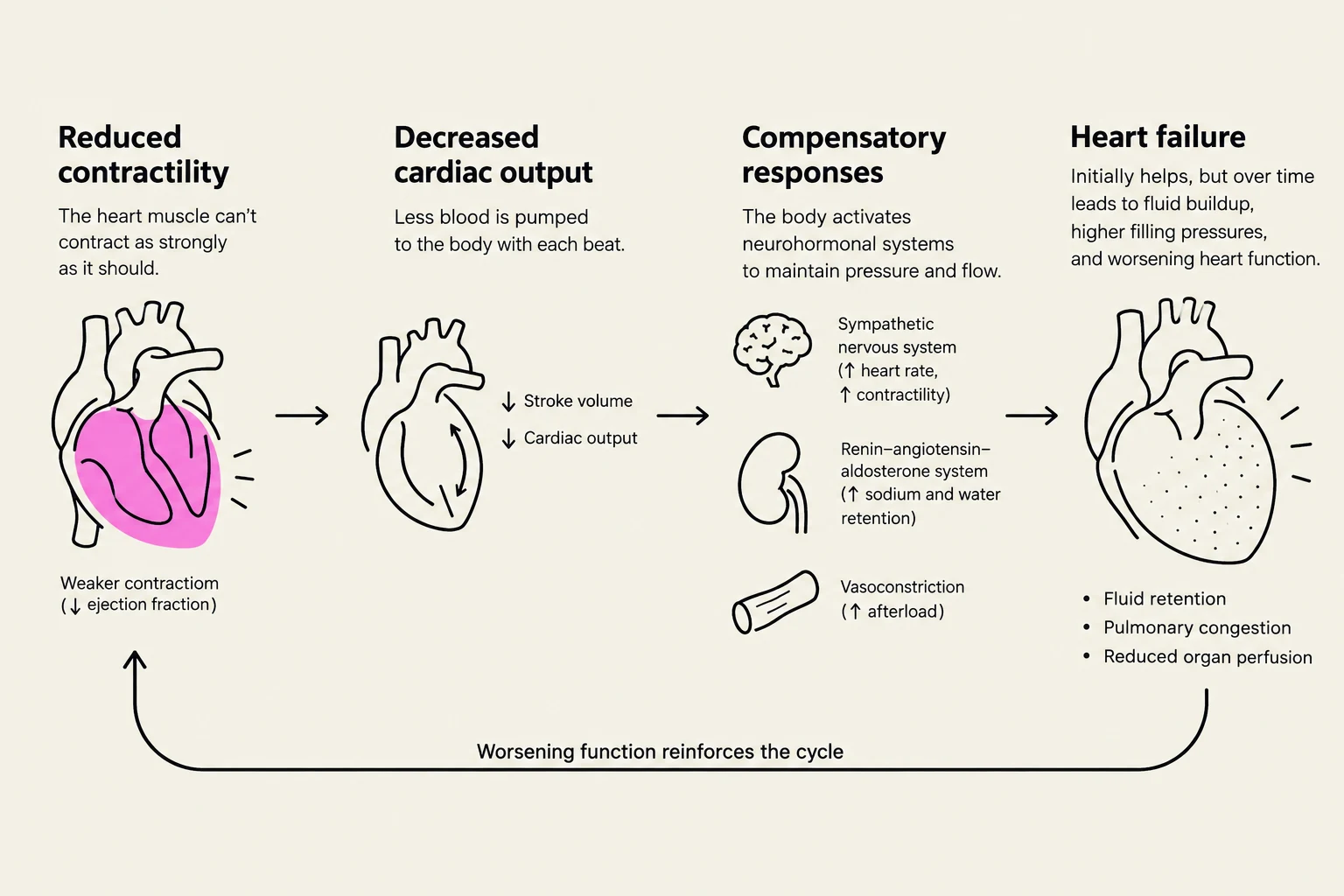
This diagram illustrates the progression of heart failure, starting with reduced contractility and decreased cardiac output, leading to compensatory responses and ultimately heart failure with symptoms like fluid retention and pulmonary congestion.
Clinical Note: While the body uses compensatory mechanisms like the Sympathetic Nervous System and the RAAS to support perfusion in the short term, these responses eventually become harmful. Chronic tachycardia increases oxygen demand, and fluid retention increases preload and afterload, creating a cycle of worsening failure.
Keep reading for free.
Create an account to finish this reading and make it part of your study routine.