A comprehensive review of cardiac anatomy, physiology, common congenital lesions, ECG patterns related to electrolyte imbalances, and a systematic approach to physical examination of the heart.
Understanding the cardiac system requires a dual focus on the mechanical flow of blood and the electrical impulses that drive it. A clinician must be able to bridge the gap between anatomical knowledge and the physical findings—such as murmurs, pulses, and ECG changes—that signal underlying pathology. This article reviews the essential components of cardiac anatomy, common congenital lesions, the impact of electrolyte imbalances on cardiac rhythm, and the systematic approach to physical examination.
Anatomy and Physiology of Cardiac Function
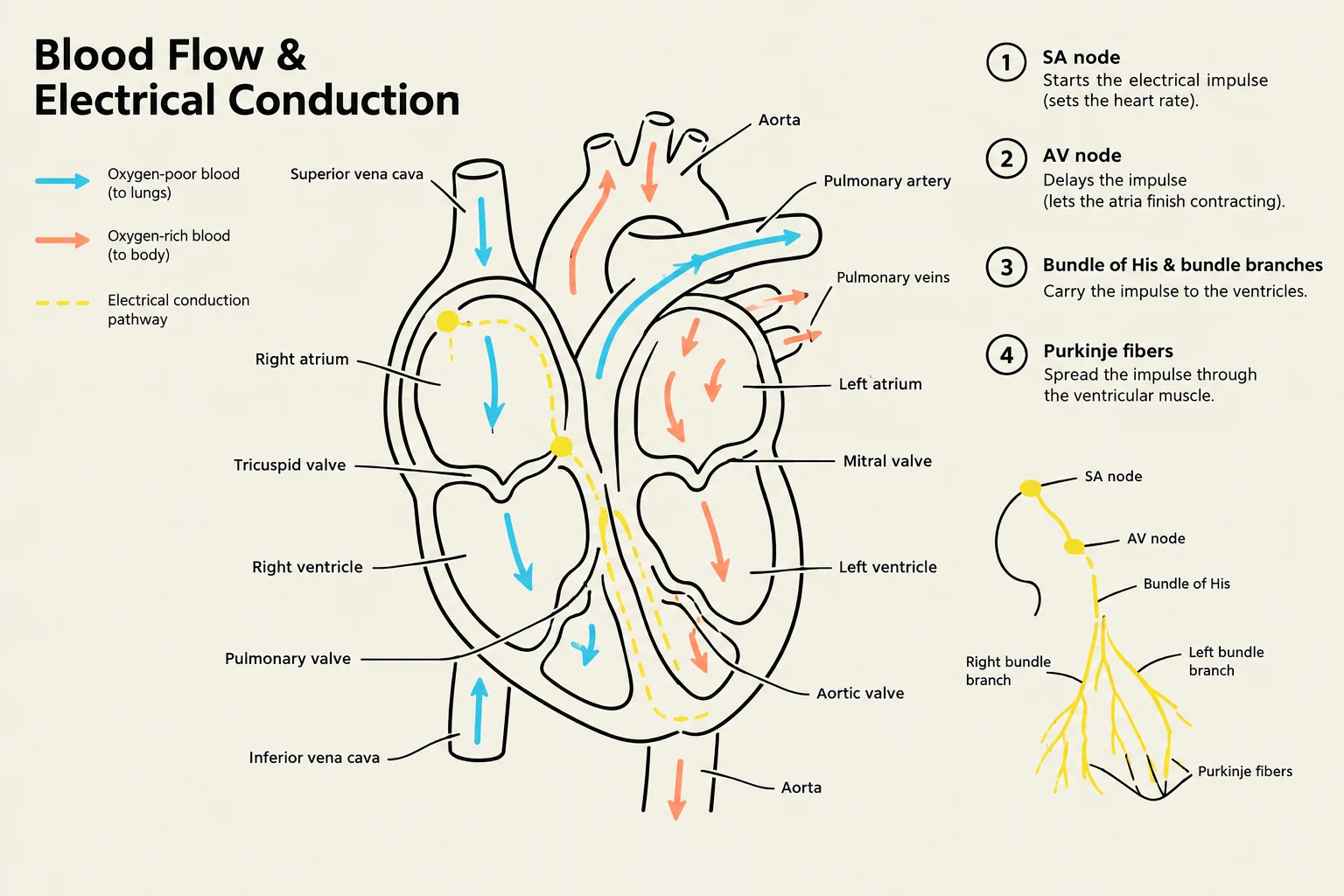
The cardiac cycle is a highly coordinated process of pressure changes and valve movements. Normal blood flow follows a specific, unidirectional pathway: the venae cavae→right atrium→tricuspid valve→right ventricle→pulmonic valve→lungs→left atrium→mitral valve→left ventricle→aortic valve→systemic circulation.
Simultaneously, the electrical conduction system ensures the ventricles contract in a manner that maximizes cardiac output. The impulse is initiated at the SA node, travels to the AV node, passes through the His bundle, descends through the bundle branches, and finally reaches the ventricular myocardium via the Purkinje fibers.
Diagram illustrating blood flow and electrical conduction through the human heart, detailing the roles of the SA node, AV node, Bundle of His, and Purkinje fibers in regulating heart rate and contraction.
Congenital Cardiac Lesions
Congenital heart defects often present with distinct hemodynamic shifts and physical exam findings. Recognizing these patterns is critical for early intervention.
Coarctation of the Aorta
This condition involves a narrowing of the aorta, typically near the ductus arteriosus. This creates a pressure gradient where the upper body experiences hypertension while the lower body receives reduced flow.
Key Finding: A radiofemoral delay, where blood pressure is higher in the arms than in the legs, and femoral pulses may be weak or delayed.
Pulmonic Stenosis
Pulmonic stenosis is an obstruction of blood flow from the right ventricle into the pulmonary artery, which increases right ventricular pressure.
Key Finding: An ejection click and a harsh crescendo-decrescendo systolic murmur heard at the left upper sternal border.
Tetralogy of Fallot
This complex lesion consists of four components: a ventricular septal defect (VSD), an overriding aorta, pulmonic stenosis, and right ventricular (RV) hypertrophy. The combination of these defects leads to right-to-left shunting, resulting in cyanosis and clubbing.
Clinical Pearl: Patients may experience hypercyanotic spells (often called "Tet spells"), which can be partially relieved by placing the patient in a squatting or knee-chest position.
Ventricular Septal Defect (VSD)
A VSD is an opening between the left and right ventricles. The clinical presentation depends heavily on the size of the defect:
Small defects: Often produce a loud, harsh holosystolic murmur at the left lower sternal border.
Large defects: May lead to significant shunting, resulting in tachypnea, diaphoresis, poor growth, and eventual heart failure.
Electrolyte Abnormalities and ECG Patterns
Electrolytes play a fundamental role in maintaining the resting membrane potential of cardiac cells. Imbalances can manifest as life-threatening changes on an electrocardiogram (ECG).
Calcium and Magnesium
Hypercalcemia: Characterized by a shortened QT interval.
Hypocalcemia: Results in a prolonged QT interval, which increases the risk of ventricular arrhythmias.
Hypomagnesemia: Also causes QT prolongation and significantly increases the risk of torsades de pointes.
Potassium
Potassium is perhaps the most critical electrolyte regarding cardiac rhythm stability.
Hyperkalemia: Presents with peaked T waves, PR prolongation, flattened P waves, and eventually QRS widening or a "sine-wave" pattern.
Hypokalemia: Presents with ST depression, flattened T waves, and the appearance of prominent U waves.
Exam Tip: When assessing an ECG, always look at the T wave and the QT interval first if you suspect potassium or calcium imbalances. Remember: "High Calcium = Short QT; Low Calcium = Long QT."
Clinical Cardiac Assessment
A systematic physical examination is essential to differentiate between physiologic and pathologic findings.
Inspection and Palpation
Begin by inspecting the patient for cyanosis, clubbing, edema, or jugular venous distention. During palpation, locate the Precordial Impulse (PMI) at the left fifth interspace near the midclavicular line. A lateral displacement of the PMI may suggest cardiomegaly.
Clinicians should also palpate for thrills—palpable vibrations that typically indicate a loud murmur (grade 4/6 or higher). Peripheral pulses should be graded on a scale of 0 to 4+:
0: Absent
1+: Weak/Thready
2+: Expected/Normal
3+: Increased
4+: Bounding
Auscultation: Heart Sounds and Murmurs
Auscultation should be performed systematically using both the diaphragm (for high-pitched sounds) and the bell (for low-pitched sounds).
Heart Sounds:
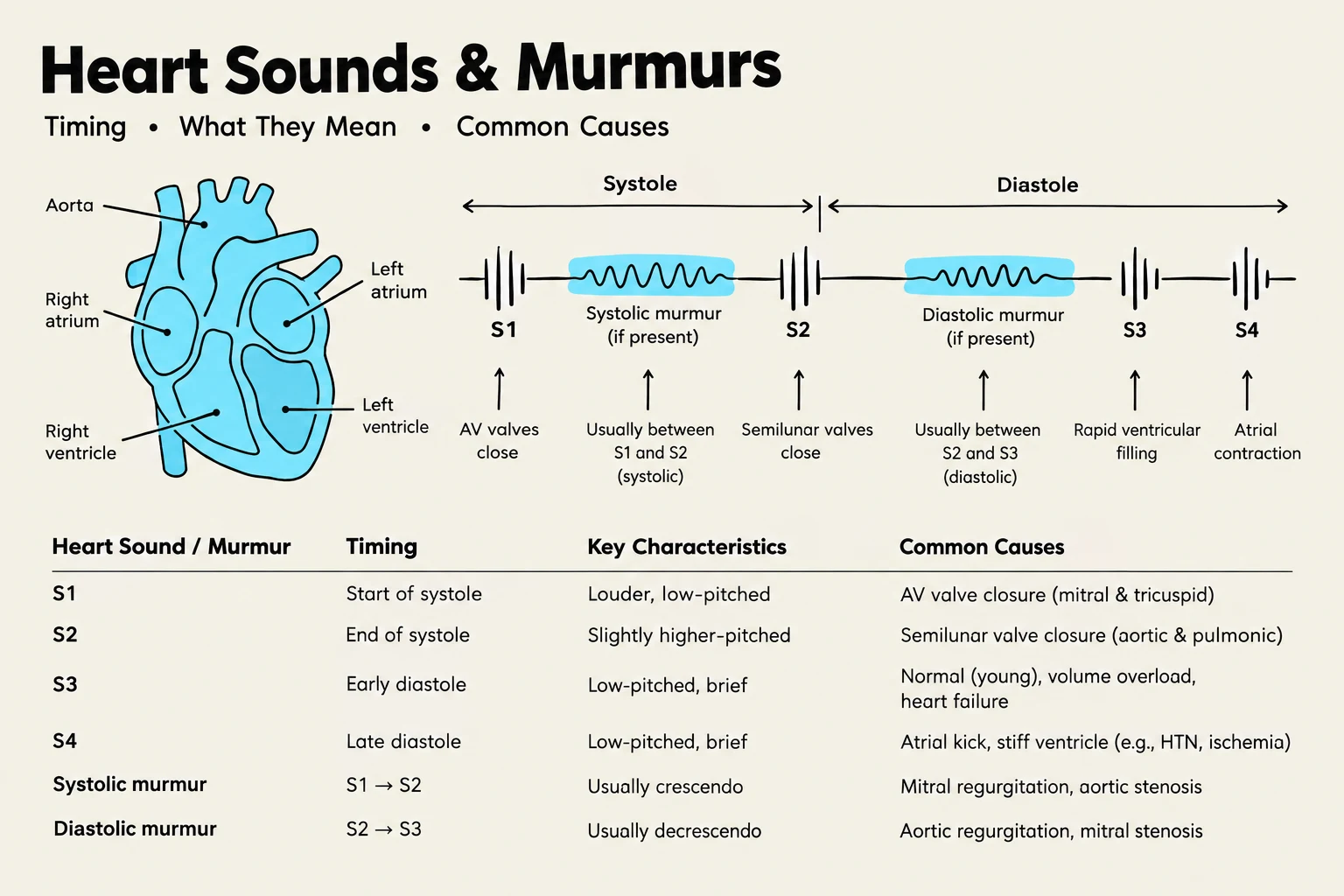
S1: Closure of the mitral and tricuspid valves; marks the beginning of systole.
S2: Closure of the aortic and pulmonic valves; marks the end of systole.
S3: A low-pitched sound following S2. While normal in children and pregnant women, in older adults, it may indicate volume overload or ventricular failure.
S4: A low-pitched sound preceding S1, indicating a stiff, noncompliant ventricle (e.g., from hypertension).
Murmurs:
Murmurs are classified by their timing in the cardiac cycle. Diastolic murmurs are almost always pathologic.
Systolic Murmurs: Can be caused by stenosis (crescendo-decrescendo) or regurgitation (holosystolic).
Aortic Regurgitation: A high-pitched, blowing, early diastolic decrescendo murmur, often heard best while the patient leans forward at end-expiration.
Pericardial Friction Rub: A scratchy, grating sound heard best with the diaphragm while the patient leans forward; this is a hallmark of pericarditis.
This diagram illustrates heart sounds and murmurs, detailing their timing, key characteristics, and common causes, alongside a simplified representation of the human heart.
Urgent Findings and Red Flags
Certain clinical presentations require immediate medical intervention to prevent adverse outcomes.
Immediate intervention is required if a patient presents with:
Chest pain accompanied by diaphoresis (sweating) or vomiting.
Syncope (fainting), especially if it occurs with exertion.
New neurologic deficits or sudden changes in mental status.
Pulseless, cold, or discolored limbs.
Unstable ventricular rhythms or signs of shock.
Keep reading for free.
Create an account to finish this reading and make it part of your study routine.