Cardiac Remodeling and Hypertrophy: Mechanisms of Adaptation and Failure
This content explains cardiac remodeling, a process where the heart changes size, shape, and function in response to stress. It details concentric hypertrophy (thickening due to pressure overload) and eccentric hypertrophy (dilation due to volume overload), and how these adaptive changes can transition to pathological
The human heart is a remarkably dynamic organ, capable of altering its structure to meet the physiological demands of the body. However, this ability to adapt is a double-edged sword. While initial structural changes can help maintain cardiac output in the face of stress, these same processes often transition from beneficial compensation to pathological destruction. This phenomenon, known as cardiac remodeling, is a central driver in the progression of heart failure.
The Concept of Cardiac Remodeling
Cardiac remodeling refers to the changes in size, shape, structure, and function of the heart throughout various stages of disease. It is not merely a change in the "size" of the heart, but a complex reorganization involving myocyte hypertrophy (increase in cell size), changes in the extracellular matrix (fibrosis), and alterations in the geometry of the ventricular chambers.
When the heart faces a chronic stressor—such as high blood pressure or a damaged muscle after a heart attack—it attempts to adapt to maintain stroke volume and cardiac output. This adaptation can be categorized into two primary morphological patterns: concentric hypertrophy and eccentric hypertrophy.
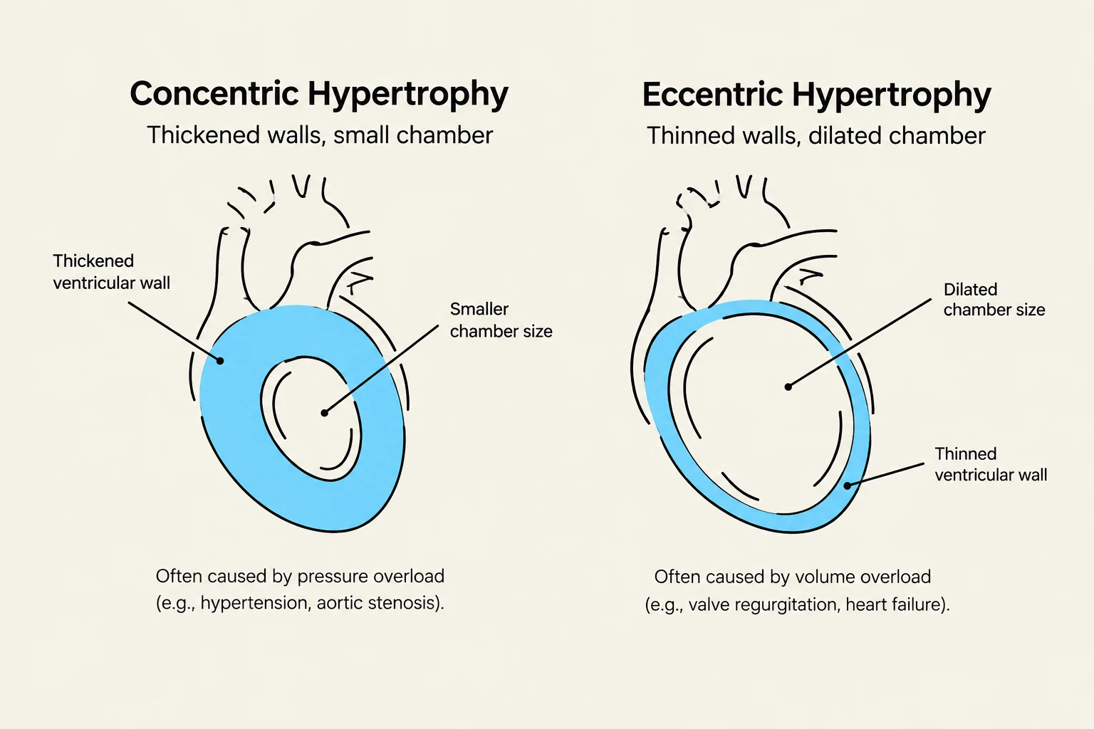
Diagrams illustrating concentric and eccentric cardiac hypertrophy, detailing thickened walls and small chamber size in concentric hypertrophy, often due to pressure overload, and thinned walls with dilated chamber size in eccentric hypertrophy, often due to volume overload.
Concentric Hypertrophy: Responding to Pressure Overload
Concentric hypertrophy occurs when the heart must pump against an increased resistance, known as afterload. A classic example is systemic hypertension (high blood pressure) or aortic stenosis (narrowing of the aortic valve).
In this scenario, the ventricular wall thickens by adding sarcomeres in parallel. This increases the thickness of the muscular wall relative to the chamber radius, which helps normalize the wall stress according to Laplace's Law.
Exam Tip: If a question mentions "increased afterload," "hypertension," or "pressure overload," think Concentric Hypertrophy. The key visual cue is a thickened wall and a narrowed ventricular lumen.
The relationship between wall stress (σ), intraventricular pressure (P), ventricular radius (r), and wall thickness (h) is defined by the Law of Laplace:
σ=2hP⋅r
By increasing the thickness (h), the heart attempts to keep the wall stress (σ) from rising to dangerous levels despite the high pressure (P). While effective in the short term, the thickened muscle eventually becomes stiff, leading to diastolic dysfunction (difficulty filling).
Eccentric Hypertrophy: Responding to Volume Overload
In contrast, eccentric hypertrophy occurs in response to volume overload. This happens when the heart must accommodate an abnormally large volume of blood, such as in valvular regurgitation (where blood leaks backward) or in athletes undergoing intense endurance training.
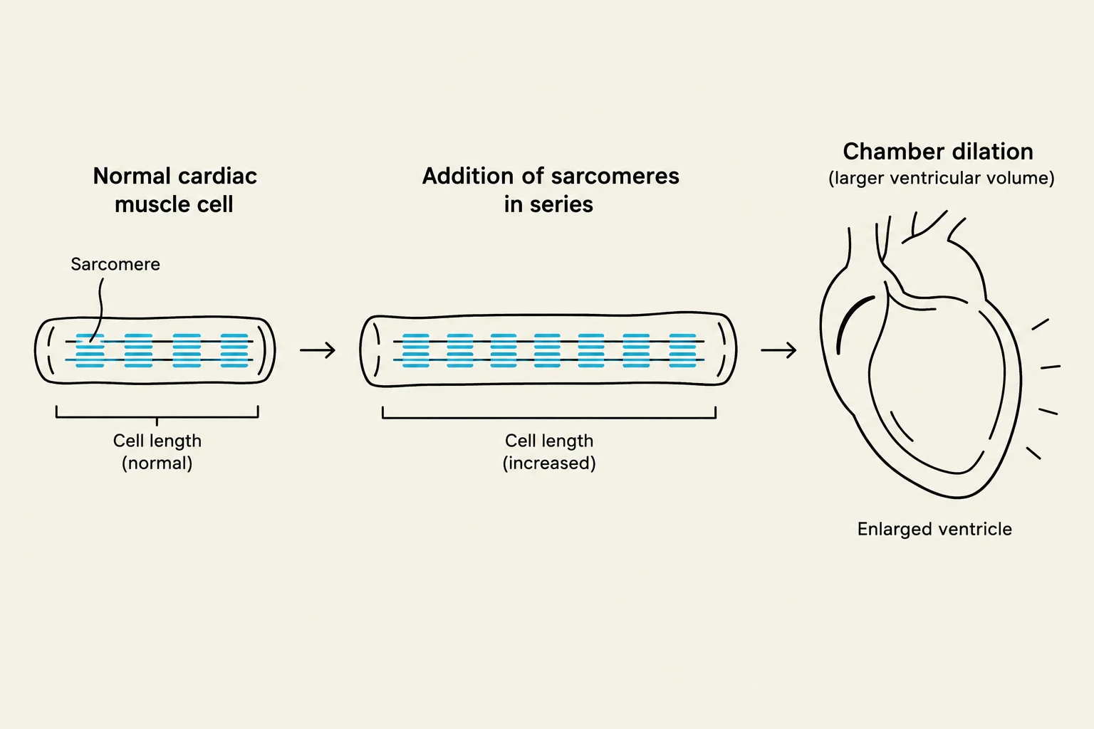
In eccentric hypertrophy, the myocytes grow by adding sarcomeres in series. This results in the lengthening of the myocytes, which causes the ventricular chamber to dilate (expand).
This diagram illustrates how the addition of sarcomeres in series can lead to chamber dilation in a cardiac muscle cell, resulting in a larger ventricular volume.
While a larger chamber can hold more blood (increasing preload), the walls often become thinner and weaker over time. This is frequently seen in the late stages of dilated cardiomyopathy.
From Compensation to Heart Failure
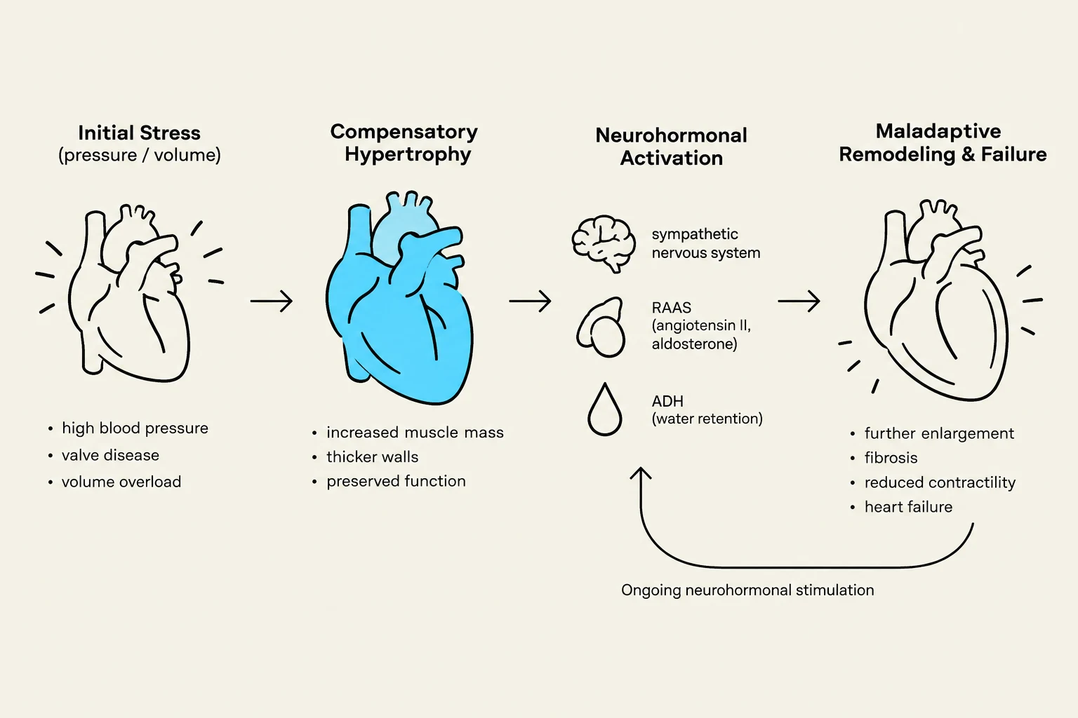
The transition from "adaptive" remodeling to "maladaptive" remodeling is the hallmark of congestive heart failure. What begins as a way to maintain cardiac output eventually becomes a cycle of decline.
Myocyte Death and Fibrosis: As myocytes undergo hypertrophy, they may outgrow their blood supply, leading to localized ischemia and cell death. This triggers the activation of fibroblasts, which lay down collagen (fibrosis), making the heart stiff and non-compliant.
Neurohormonal Activation: The body senses decreased cardiac output and activates the Renin-Angiotensin-Aldosterone System (RAAS) and the sympathetic nervous system. While intended to increase blood pressure and volume, these systems actually increase afterload and promote further pathological remodeling.
Chamber Dilation and Wall Thinning: In many cases, the heart eventually loses its ability to maintain wall tension, leading to massive dilation and a significant drop in ejection fraction.
This diagram illustrates the progression of heart conditions from initial stress to maladaptive remodeling and failure, detailing compensatory hypertrophy and neurohormonal activation.
Summary Table: Comparison of Hypertrophic Patterns
Feature
Concentric Hypertrophy
Eccentric Hypertrophy
Primary Stimulus
Pressure Overload (Afterload)
Volume Overload (Preload)
Sarcomere Addition
In Parallel (Thickening)
In Series (Lengthening)
Chamber Size
Decreased/Normal
Increased (Dilation)
Wall Thickness
Increased
Normal or Decreased
Common Example
Hypertension, Aortic Stenosis
Mitral Regurgitation, Dilated Cardiomyopathy
Understanding these mechanisms is vital for clinical management, as treatment often focuses on reducing the very stressors—afterload and preload—that drive this remodeling process.
Keep reading for free.
Create an account to finish this reading and make it part of your study routine.