Cardiac Physiology: Electrical and Mechanical Function
Explore the fundamental principles of cardiac physiology, including the cellular basis of heart function, electrical activity (action potentials), intrinsic and extrinsic innervation, and the mechanical phases of the cardiac cycle.
The human heart is a sophisticated biological pump that relies on a seamless integration of electrical signals and mechanical actions. To understand how the heart functions, one must look at the interplay between specialized cells, the electrical impulses they generate, and the pressure gradients that drive blood flow through the body. This article explores the cellular foundations, the intrinsic and extrinsic control systems, and the mechanical phases of the cardiac cycle.
The Cellular Foundation of the Heart
The heart is composed of several distinct cell types that work in concert to ensure efficient circulation. While endocardial cells line the inner surfaces and valves of the heart, the primary drivers of cardiac function are the myocardial and conduction cells.
Myocardial Cells
Myocardial cells, or contractile cells, are responsible for the actual kinetic work of the heart. They are found in the atrial walls and the myocardial layer of the ventricles. These cells contain myofibrils, which are made of even smaller units called sarcomeres. Within these sarcomeres, the proteins actin and myosin slide past one another to facilitate contraction.
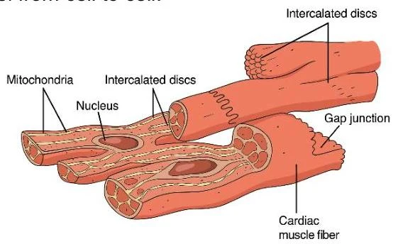
Diagram illustrating the structure of cardiac muscle fibers, highlighting key components such as mitochondria, nucleus, intercalated discs, and gap junctions.
A critical feature of myocardial cells is the presence of intercalated discs. These discs contain desmosomes to anchor fibers together and gap junctions that act as gateways, allowing electrical impulses to travel rapidly from cell to cell.
Conduction Cells
Unlike myocardial cells, conduction cells (also known as auto-arrhythmic cells) are specialized to initiate and transmit electrical impulses. While they do not have the ability to contract, they are the "electrical wiring" that tells the myocardial cells when to squeeze.
Electrical Activity: Action Potentials
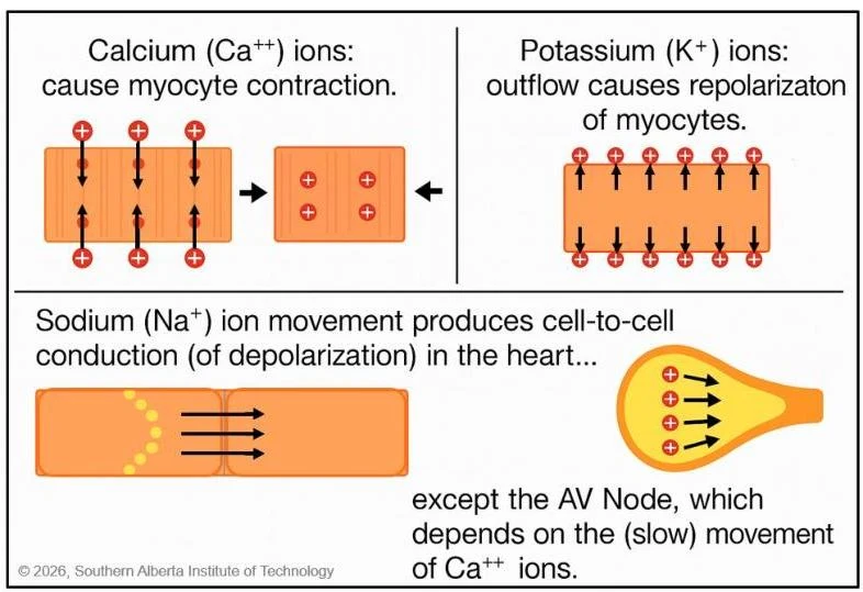
The electrical signal that triggers a heartbeat is known as an action potential. This is a wave of electrical discharge caused by the movement of ions—specifically Sodium (Na+), Potassium (K+), and Calcium (Ca++)—across the cell membrane via electrochemical gradients.
The cycle of an action potential consists of two main phases:
Depolarization: The cell moves from a negative resting state (polarized) to a positive state. In the heart, depolarization is the trigger for myocardial contraction.
Repolarization: The cell returns to its negative resting state.
This diagram illustrates the roles of calcium, potassium, and sodium ions in myocyte contraction and conduction, with a note on the AV node's dependence on calcium ions.
Clinical Note: Because these electrical changes depend on ion movement, the heart's function can be significantly impacted by dietary sodium and potassium levels or calcium concentrations in the bloodstream.
Intrinsic Innervation: The Conduction Pathway
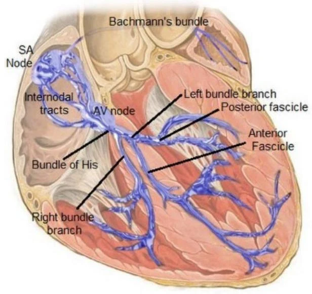
Intrinsic innervation refers to the heart's internal electrical conduction system, which allows it to generate its own rhythm. This journey begins at the natural pacemaker and follows a specific sequence:
SA Node (Sinoatrial Node): Located in the upper wall of the right atrium, this is the dominant pacemaker. It initiates a wave of depolarization that creates a "sinus rhythm" of 60–100 beats per minute (bpm).
AV Node (Atrioventricular Node): The only pathway between the atria and ventricles. It slows the depolarization wave, allowing time for the atria to contract and fill the ventricles before ventricular contraction begins. It can act as a backup pacemaker at 40–60 bpm.
Bundle of HIS: Located distal to the AV node, the signal speeds up here as it enters the bundle branches.
Bundle Branches (Right and Left): These carry the impulse toward the apex of the heart.
Purkinje Fibers: These individual fibers deliver the impulse to the ventricular muscle cells. They can generate a backup rate of 30–40 bpm.
Diagram of the cardiac conduction system, showing the SA node, AV node, Bundle of His, and bundle branches.
Extrinsic Innervation: External Control
While the heart can beat on its own, the nervous system provides extrinsic innervation to adjust the heart rate and strength of contraction based on the body's needs.
Sympathetic Nervous System: The "fight or flight" system. It releases norepinephrine to increase both heart rate (chronotropy) and the force of contraction (inotropy).
Parasympathetic Nervous System: The "rest and digest" system. It acts as a brake via the vagus nerve, releasing acetylcholine to slow the heart rate.
Pressure Gradients and the Cardiac Cycle
Blood flow is not random; it is entirely dependent on pressure gradients. Blood always moves from an area of higher pressure to an area of lower pressure. When the heart contracts (depolarization), pressure in that chamber rises; when it relaxes (repolarization), pressure falls.
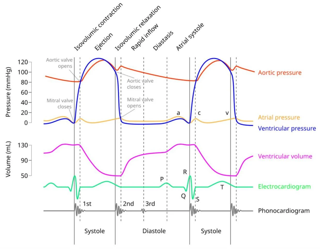
The cardiac cycle describes the complete sequence of mechanical and electrical events in one heartbeat. It is often analyzed using a Wiggers diagram, which integrates ECG, pressure, and volume data.
This diagram illustrates the pressure and volume changes in the heart chambers during the cardiac cycle, alongside the electrocardiogram and phonocardiogram.
The Four Phases of the Cardiac Cycle
The cycle can be broken down into four primary functional periods:
Isovolumic Contraction Time (IVCT): Part of systole. The ventricles begin to contract, but all valves (mitral/tricuspid and aortic/pulmonic) are closed. Pressure rises sharply, but volume remains constant.
Systole (The "Big Squeeze"): The period where the ventricles eject blood. This begins when ventricular pressure exceeds aortic pressure, forcing the semilunar valves open.
Isovolumic Relaxation Time (IVRT): Part of diastole. After the semilunar valves close, the ventricles relax. All valves are closed again, and pressure falls rapidly while volume remains the same.
Diastole (Ventricular Filling): The period where the ventricles fill with blood. This includes:
Early Filling: Rapid blood flow into the ventricles as pressure drops.
Diastasis: A slower filling phase where pressures equalize.
Atrial Kick: The final 30% of filling caused by atrial contraction.
Translating Electricity to the ECG
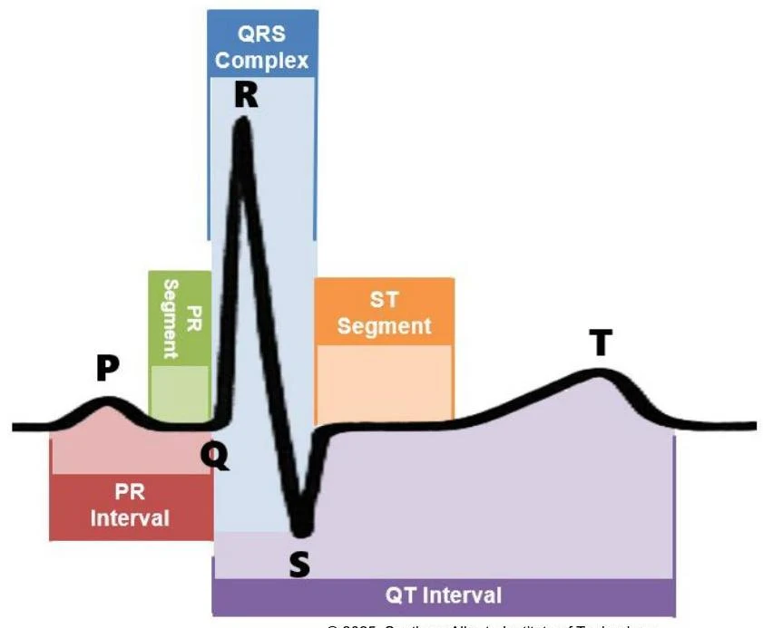
An Electrocardiogram (ECG) records the electrical activity of the heart via electrodes on the skin. Each wave on the ECG corresponds to a specific electrical event:
P Wave: Atrial depolarization.
QRS Complex: Ventricular depolarization (the large spike).
T Wave: Ventricular repolarization.
An electrocardiogram (ECG) waveform illustrating different components such as the P wave, QRS complex, T wave, and intervals like the PR interval and QT interval.
Study Tip: Remember that the PR interval includes both the electrical event (P wave) and the mechanical event (atrial contraction), whereas a segment (like the PR segment) refers only to the electrical pause between waves.
Keep reading for free.
Create an account to finish this reading and make it part of your study routine.