The heart is a sophisticated muscular pump responsible for circulating blood throughout the body, ensuring that oxygen and nutrients reach tissues while metabolic waste products are removed. Understanding the heart requires a dual approach: studying its physical anatomy (the "hardware") and its electrical and mechanical processes (the "software").
Anatomy of the Heart
The heart is divided into four distinct chambers: the right and left atria (upper chambers) and the right and left ventricles (lower chambers). A muscular wall known as the septum divides the heart vertically, separating the left and right sides to prevent the mixing of oxygenated and deoxygenated blood.
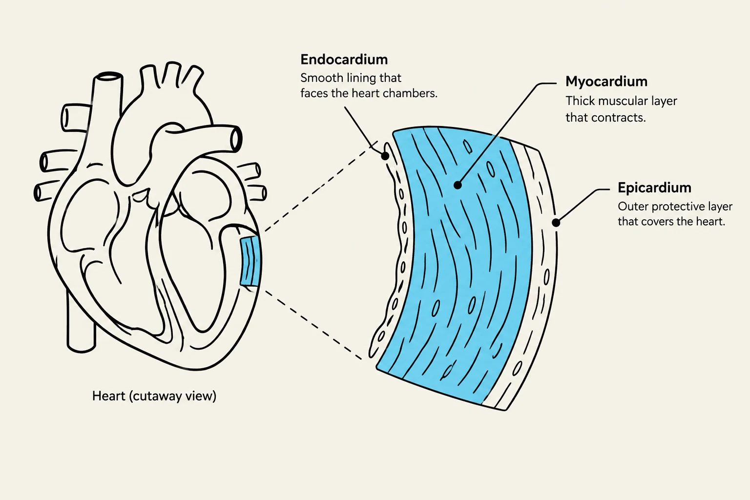
The heart wall itself is composed of three specialized layers:
Endocardium: The innermost layer that lines the chambers.
Myocardium: The middle layer, consisting of the actual cardiac muscle tissue responsible for contraction.
Epicardium: The outermost layer, often referred to as the heart sac.
Diagram illustrating the layers of the heart wall: endocardium, myocardium, and epicardium.
Blood Flow and Valvular Function
Blood moves through the heart in a specific, one-way circuit. Veins carry blood back to the heart, while arteries carry blood away from it.
The Circulatory Path
The flow of deoxygenated blood begins as it enters the Right Atrium via the Superior and Inferior Vena Cava. It passes through the Tricuspid Valve into the Right Ventricle, then through the Pulmonary Semilunar Valve into the Pulmonary Artery, which leads to the lungs for oxygenation. Once oxygenated, the blood returns via the Pulmonary Vein to the Left Atrium. It then moves through the Mitral Valve into the Left Ventricle, and is finally pumped through the Aortic Semilunar Valve into the Aorta to be distributed to the rest of the body.
The Role of Valves
Valves act as one-way gates, ensuring that blood flows in the correct direction and preventing backflow. They are categorized into two types:
Atrioventricular (AV) Valves: These sit between the atria and ventricles. They include the Mitral valve (between the left atrium and left ventricle) and the Tricuspid valve (between the right atrium and right ventricle).
Semilunar Valves: These sit at the exits of the ventricles. They include the Aortic valve and the Pulmonic valve.
Clinical Note: Heart Sounds
When listening to a patient's heart, the "Lub-Dub" sounds are produced by the closing of these valves.
S1 (Lub): Caused by the closure of the Mitral and Tricuspid valves.
S2 (Dub): Caused by the closure of the Aortic and Pulmonic valves.
Electrical Conduction and Innervation
The heart does not rely on the brain for every single beat; it has its own intrinsic electrical system that coordinates contraction.
The Conduction Pathway
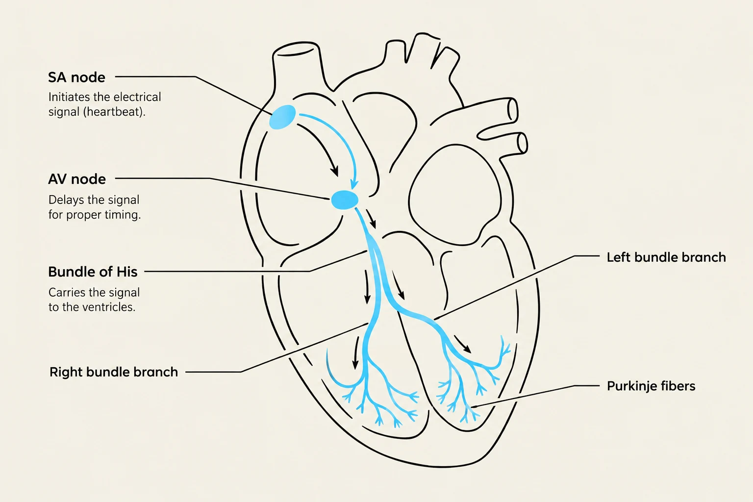
The electrical impulse follows a specific route:
SA Node (Sinoatrial Node): The natural pacemaker.
AV Node (Atrioventricular Node): Delays the impulse to allow ventricles to fill.
Bundle of His: Transmits the impulse to the ventricles.
Left and Right Bundle Branches: Carries the impulse down the septum.
Purkinje Fibers: Distributes the impulse throughout the ventricular myocardium to trigger contraction.
Diagram illustrating the electrical conduction system of the heart, including the SA node, AV node, Bundle of His, bundle branches, and Purkinje fibers.
Autonomic Regulation
While the heart is self-exciting, the autonomic nervous system modulates its activity:
Sympathetic Nervous System (SNS): Acts like an accelerator, increasing heart rate, conduction speed, and the strength of contractions.
Parasympathetic Nervous System (PNS): Acts like a brake, slowing the heart rate, slowing conduction, and decreasing the strength of contractions.
The Cardiac Cycle and Hemodynamics
The cardiac cycle refers to one complete heartbeat, consisting of contraction and relaxation.
Systole and Diastole
Systole: The phase where the ventricles contract and eject blood into the arteries.
Diastole: The phase where the ventricles relax and fill with blood.
Measuring Cardiac Performance
To understand how well the heart is working, we look at several hemodynamic parameters:
Cardiac Output (CO) is the volume of blood pumped by the heart per minute. It is heavily influenced by Stroke Volume (SV)—the amount of blood ejected in a single contraction.
Three primary factors affect Stroke Volume:
Preload: The degree of stretch in the ventricular muscle at the end of diastole (the "filling" pressure).
Afterload: The resistance the heart must pump against to eject blood (often related to blood pressure).
Contractility: The innate strength of the heart muscle's contraction.
Blood Pressure and Perfusion
Blood pressure is the measurement of pressure exerted by blood against the arterial walls.
Pulse Pressure: The difference between Systolic Blood Pressure (SBP) and Diastolic Blood Pressure (DBP).
Mean Arterial Pressure (MAP): This represents the average perfusion pressure felt by the organs. It is a critical indicator of whether vital organs are receiving enough blood.
The formula for MAP is:
MAP = \frac{SBP + 2(DBP)}{3}
\end{aligned}$$
> **Critical Value:** A MAP greater than 60 mmHg is generally necessary to sustain vital organ function.
## Coronary Circulation and Aging
### Coronary Blood Flow
The heart muscle itself requires a constant supply of oxygenated blood, provided by the coronary arteries. The **Left Coronary Artery** supplies the left atrium, left ventricle, and the septum, while the **Right Coronary Artery** supplies the SA node, AV node, right atrium, and right ventricle.
The heart has a protective mechanism: **Collateral Arteries**. These are connections between different branches of arteries that can develop to provide alternative routes for blood flow if a primary vessel becomes occluded (blocked).
### Age-Related Changes
As humans age, several physiological changes occur in the cardiovascular system:
* Decreased myocardial contractility.
* Changes in valve structure.
* Changes in the SA node and nerve fiber conduction.
* Increased risk of dysrhythmias (irregular rhythms).
* Arterial and venous changes, often leading to increased pulse pressure or hypertension (HTN).
* Prolonged recovery times and slower adaptation to physiological stress.
$$$
$$$
Keep reading for free.
Create an account to finish this reading and make it part of your study routine.