Cardiac Hemodynamics and Cardiovascular Assessment
An overview of cardiac hemodynamics, including principles of blood flow, cardiac output regulation, and methods for assessing cardiovascular health. Covers heart function, blood pressure, and common cardiovascular symptoms.
Understanding how the heart functions as a pump and how to assess its efficiency is fundamental to clinical practice. Cardiovascular health relies on the seamless integration of electrical signals, mechanical contractions, and the physics of fluid dynamics. When these systems fail, patients may present with life-threatening conditions ranging from myocardial ischemia to hypertensive emergencies.
Principles of Cardiac Hemodynamics
Cardiac hemodynamics is governed by the fundamental principle that fluid moves from areas of higher pressure to areas of lower pressure. The heart operates through a rhythmic cycle consisting of two primary phases: diastole and systole.
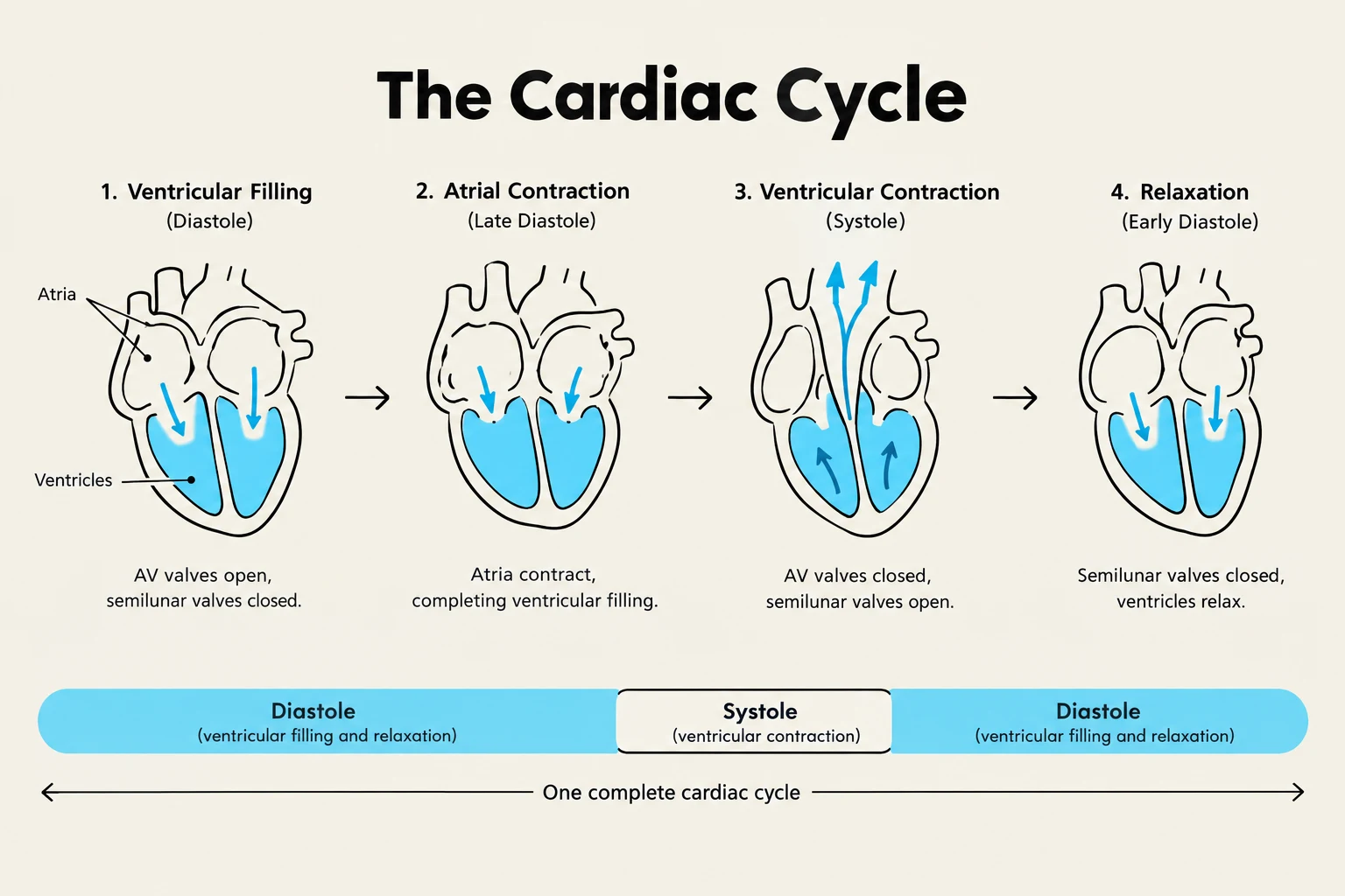
During diastole, the heart muscle relaxes, allowing the chambers to fill with blood. This phase is critical for myocardial perfusion; the coronary arteries are primarily perfused during diastole. In contrast, systole involves the contraction of the atria (atrial systole) followed by the powerful contraction of the ventricles (ventricular systole), which ejects blood into the pulmonary artery and the aorta.
The cardiac cycle illustrates the sequence of events in the heart during one complete heartbeat, encompassing diastole (ventricular filling and relaxation) and systole (ventricular contraction).
Cardiac Output and Stroke Volume Regulation
Cardiac Output (CO) is the total volume of blood ejected by the ventricles per minute. For a resting adult, a normal CO is typically between 4 to 6 L/min. It is calculated using the following relationship:
CO=HR×SV
Where Heart Rate (HR) is the number of beats per minute and Stroke Volume (SV) is the volume of blood ejected per single beat.
Stroke volume is not a static number; it is dynamically regulated by three key physiological factors:
Preload: This is the degree of ventricular stretch at the end of diastole. According to the Frank-Starling Law, an increase in preload (e.g., through IV fluids or blood transfusions) leads to an increase in stroke volume. Conversely, dehydration or blood loss decreases preload and SV.
Afterload: This represents the resistance against which the ventricles must pump to eject blood. Increased afterload, such as that caused by systemic vasoconstriction or hypertension, decreases stroke volume.
Contractility: This refers to the intrinsic force of the myocardial contraction. Increased contractility increases SV, while decreased contractility (often seen in heart failure) decreases SV.
To assess the efficiency of the left ventricle, clinicians use the Ejection Fraction (EF). This is the percentage of the end-diastolic blood volume that is ejected with each beat. A normal EF is considered to be >55%, while an EF <40% indicates decreased left ventricular function.
Autonomic and Baroreceptor Regulation
The body maintains cardiovascular homeostasis through complex feedback loops. The Autonomic Nervous System (ANS) is the primary regulator of heart rate and cardiac output:
Sympathetic Nervous System: Stimulates beta-1 receptors in the SA node to increase heart rate and contractility.
Parasympathetic Nervous System: Mediated via the vagus nerve, this system decreases the heart rate.
Additionally, baroreceptors located in the aortic arch and carotid arteries act as pressure sensors. If blood pressure (BP) rises, baroreceptors signal the brain to decrease the heart rate; if BP drops, they trigger an increase in heart rate and vasoconstriction to restore pressure.
Cardiovascular Health History and Angina
A thorough cardiovascular assessment begins with a subjective health history to identify symptoms and risk factors. One of the most critical symptoms to assess is Angina Pectoris, which is chest pain caused by myocardial ischemia (an imbalance where oxygen demand exceeds oxygen supply).
Angina is often described as uncomfortable pressure, squeezing, or fullness in the substernal area. It may radiate to the jaw, neck, shoulders, or arms.
Clinical Tip: Distinguishing Angina Types
Stable Angina: Predictable; triggered by exertion; relieved by rest or nitroglycerin.
Unstable Angina (ACS): Unpredictable; occurs at rest; increasing in frequency/severity; requires urgent medical intervention.
Physical Assessment: Perfusion and Blood Pressure
The physical examination aims to evaluate heart function, circulating volume, and tissue perfusion.
Peripheral Circulation
Assessment of peripheral circulation involves checking both arterial and venous status:
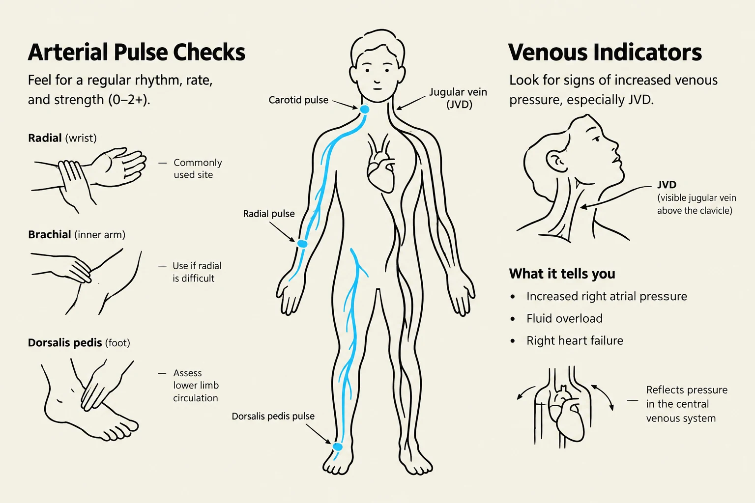
Arterial Assessment: Evaluate the rate, rhythm, and amplitude of pulses. A critical emergency to watch for is Acute Arterial Obstruction, identified by the "Six Ps": Pain, pallor, pulselessness, paresthesia, poikilothermia (coldness), and paralysis.
Venous Assessment: Look for signs of fluid overload, such as Jugular Venous Distention (JVD) or peripheral edema (swelling in the feet, ankles, or sacral area).
This illustration shows how to check for arterial pulses at various locations on the body, including the carotid, radial, and dorsalis pedis pulses. It also illustrates venous indicators, such as jugular venous distension (JVD), and what these indicators can signify regarding cardiovascular health.
Blood Pressure and Orthostatic Changes
Blood pressure is a product of cardiac output and peripheral resistance. Clinicians must also monitor Pulse Pressure, which is the difference between systolic and diastolic BP.
A significant clinical finding is Orthostatic Hypotension, defined as a drop in systolic blood pressure ≥20 mmHg or a drop in diastolic blood pressure ≥10 mmHg within three minutes of standing.
Gerontologic Considerations
Aging introduces several physiological changes that impact cardiovascular assessment:
Decreased Cardiac Output: Resulting from a slower heart rate, myocardial hypertrophy, and the stiffening of heart valves.
Atypical Symptoms: Older adults may not present with classic chest pain; instead, they may present with dyspnea (shortness of breath) or even "silent" ischemia without any reported pain.
Medication Sensitivity: There is an increased risk for adverse reactions to cardiac medications; therefore, the clinical mantra is "start low and go slow".
Keep reading for free.
Create an account to finish this reading and make it part of your study routine.