Cardiac Anatomy, Physiology, and Coronary Artery Disease
An overview of the cardiovascular system, detailing cardiac anatomy, blood flow, coronary circulation, autonomic regulation, and the pathophysiology of coronary artery disease (CAD) and atherosclerosis.
The cardiovascular system is a complex, highly regulated network designed to transport oxygen, nutrients, and waste products throughout the body. Understanding the intricate relationship between cardiac anatomy, autonomic regulation, and the progression of coronary artery disease (CAD) is essential for clinical practice. CAD remains the leading cause of death in the United States, accounting for one in four deaths. This article explores the physiological foundations of the heart and the pathological processes that lead to life-threatening cardiac events.
Cardiac Anatomy and Blood Flow
The heart operates as a dual-pump system, facilitating a specific sequence of blood flow to ensure oxygenation and systemic perfusion.
The Right Side (Pulmonary Circulation):
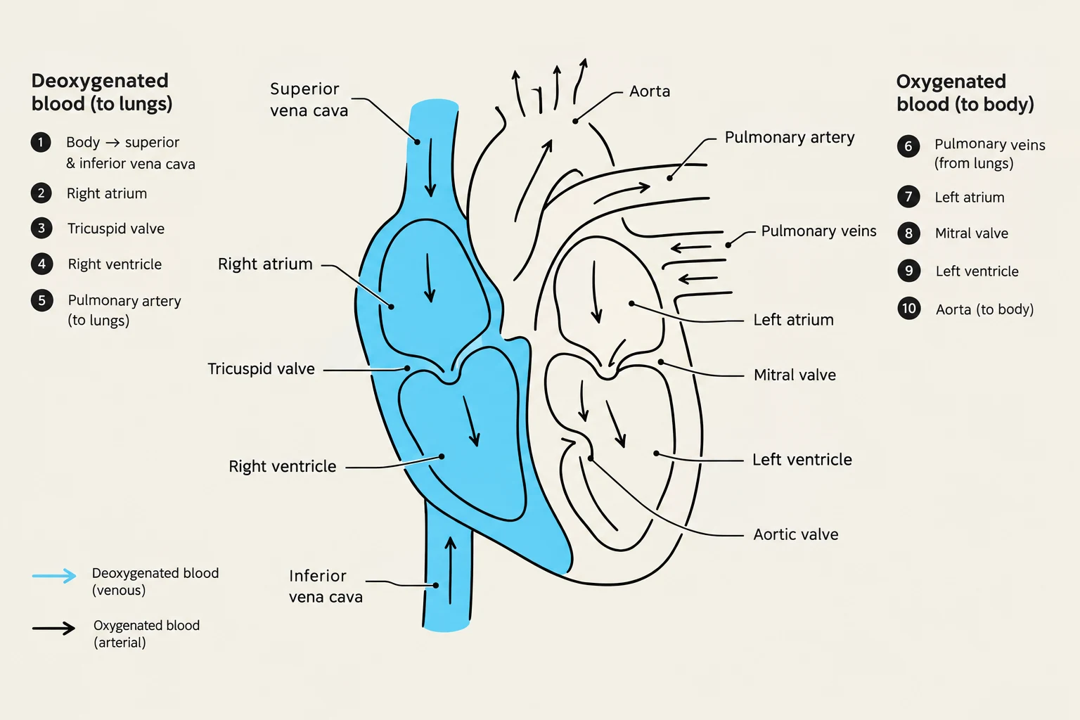
Venous blood, which is low in oxygen, enters the right atrium via the superior vena cava, inferior vena cava, and the coronary sinus. It then passes through the tricuspid valve into the right ventricle. Upon contraction, the right ventricle pumps blood through the pulmonic valve into the pulmonary artery, which carries it to the lungs for gas exchange.
The Left Side (Systemic Circulation):
Oxygenated blood returns from the lungs via the pulmonary veins to the left atrium. It moves through the mitral valve into the left ventricle, the heart's most powerful chamber. During contraction, the left ventricle ejects blood through the aortic valve into the aorta, distributing oxygen-rich blood to the rest of the body.
Diagram illustrating the flow of deoxygenated and oxygenated blood through the human heart, including key chambers and valves.
Coronary Circulation
While the heart pumps blood to the body, the myocardium (heart muscle) requires its own dedicated blood supply to function. This is provided by the coronary circulation. Crucially, most coronary blood flow occurs during diastole, the period when the myocardium is relaxed.
Left Coronary Artery: This artery divides into the left anterior descending (LAD) and the left circumflex artery. These branches supply the left atrium, the left ventricle, and the intraventricular septum.
Right Coronary Artery: This artery supplies the right atrium, the right ventricle, and a portion of the posterior wall of the left ventricle. In approximately 90% of individuals, the right coronary artery provides the blood supply to the AV node and the bundle of His. Consequently, an obstruction in this artery can lead to severe cardiac conduction defects.
Autonomic and Reflex Regulation
The heart does not beat in isolation; its rate and strength are constantly adjusted by the autonomic nervous system (ANS) to meet the body's metabolic demands.
The Autonomic Nervous System
The ANS is divided into two opposing branches:
Sympathetic Nervous System: Often called the "fight or flight" system, stimulation increases heart rate, the speed of impulse conduction through the AV node, and the force of myocardial contraction. It also stimulates alpha-1 adrenergic receptors in vascular smooth muscle, causing vasoconstriction and increasing blood pressure.
Parasympathetic Nervous System: Mediated by the vagus nerve, this system acts as a "brake," decreasing the heart rate by slowing impulses from the SA node and through the AV node.
Reflex Regulation: Baroreceptors and Chemoreceptors
The body utilizes specialized sensors to maintain homeostasis:
Baroreceptors: Located in the aortic arch and the carotid sinus, these receptors detect changes in arterial stretch (pressure). If blood pressure is too high (e.g., due to volume overload), baroreceptors trigger the vasomotor center in the brain to inhibit the sympathetic system and enhance the parasympathetic system, resulting in vasodilation and a decreased heart rate.
Chemoreceptors: Located in the aortic/carotid bodies and the medulla, these respond to chemical changes in the blood. They trigger an increase in blood pressure and respiratory rate in response to:
Hypercapnia (increased CO2)
Acidosis (decreased pH)
Hypoxemia (decreased O2)
This diagram illustrates the baroreceptor reflex, a feedback mechanism that helps regulate blood pressure by signaling the medulla oblongata in the brainstem.
Cardiac Function and Auscultation
To understand heart sounds, one must distinguish between the two phases of the cardiac cycle: systole (contraction/ejection) and diastole (relaxation/filling).
Heart Sounds
Clinicians use auscultation to listen for specific sounds that indicate valve function and ventricular health:
S1 ("lub"): The first heart sound marks the beginning of systole. It is caused by the closure of the tricuspid and mitral valves.
S2 ("dub"): The second heart sound marks the beginning of diastole. It is caused by the closure of the aortic and pulmonic valves.
S3 (Ventricular Gallop): An abnormal sound heard early in diastole. It may indicate volume overload (too much fluid), hypertension, or left ventricular dysfunction.
S4 (Atrial Gallop): An abnormal sound heard in late diastole. It is caused by the atria contracting against a resistant, often hypertrophied, ventricle.
Murmurs: These are turbulent sounds heard between S1 and S2, typically indicating abnormal blood flow patterns caused by valve disorders.
Coronary Artery Disease (CAD) and Atherosclerosis
Coronary Artery Disease is a progressive condition characterized by the narrowing or occlusion of the coronary arteries. This process is driven by atherosclerosis, the abnormal accumulation of lipids (fats) and fibrous tissue within the arterial walls.
The Stages of Atherosclerosis
Atherosclerosis does not occur overnight; it develops over many years through several distinct stages:
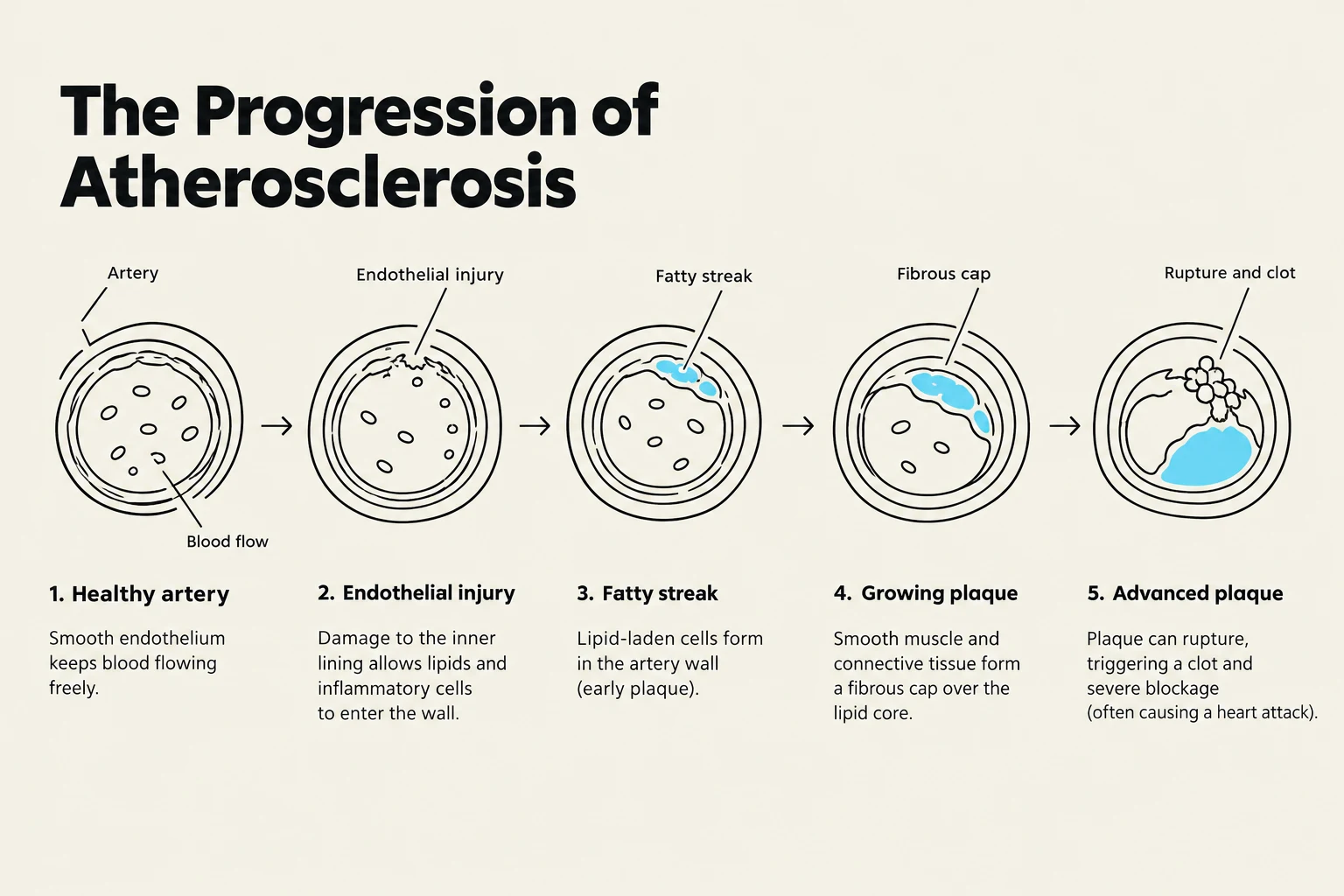
Endothelial Injury: The process begins when the inner lining of the vessel (endothelium) is damaged by factors such as smoking, high cholesterol, hypertension, diabetes, or infection.
Fatty Streak: This is the earliest lesion, characterized by lipid-filled smooth muscle cells that appear as a yellow tinge within the artery. These can be seen as early as age 18.
Fibrous Plaque: As lipoproteins (carriers of cholesterol) transport lipids into the arterial intima, collagen covers the fatty streak, forming a grayish or whitish plaque. This narrows the vessel lumen, reducing blood flow to distal tissues.
Complicated Lesion: This is the most dangerous stage. Inflammation or plaque instability leads to ulceration or rupture, causing platelets to accumulate and form a thrombus (clot). This can lead to total occlusion of the artery.
Diagram illustrating the progression of atherosclerosis, from a healthy artery to an advanced plaque with rupture and clot.
Exam Tip: Ischemia vs. Infarction
Remember that ischemia occurs when blood flow is reduced (often when a vessel is ≥50% blocked), causing oxygen deprivation. If ischemia is prolonged and not reversed, it can lead to myocardial infarction (MI). Heart cells typically become hypoxic within 10 seconds of total occlusion and can remain viable for approximately 20 minutes before irreversible damage begins.
Clinical Manifestations: Angina
When the demand for myocardial oxygen exceeds the supply provided by the coronary arteries, ischemia occurs, manifesting clinically as angina (chest pain).
Stable Angina: Usually precipitated by physical exertion.
Unstable Angina: A more serious condition characterized by new-onset angina that occurs at rest or with minimal exertion, and which increases in frequency, duration, or severity.
Commonly, angina is felt as a substernal sensation that may radiate to the neck, jaw, shoulders, or arms. Patients may also report indigestion or a burning sensation in the epigastric region.
Keep reading for free.
Create an account to finish this reading and make it part of your study routine.