An overview of cancer, covering normal cell dynamics versus cancerous growth, the genetic drivers of malignancy, stages of development from initiation to metastasis, clinical diagnosis and staging using the TNM system, and treatment modalities including palliative care.
Understanding cancer requires a deep dive into how normal cellular processes go wrong. Cancer is not a single disease but a complex collection of disorders characterized by uncontrolled cell growth, the ability to invade surrounding tissues, and the potential to spread to distant organs. This guide breaks down the journey from a single healthy cell to a metastatic malignancy, covering the genetic drivers, the stages of development, and the clinical methods used to diagnose and treat the disease.
Normal Cell Dynamics vs. Cancerous Growth
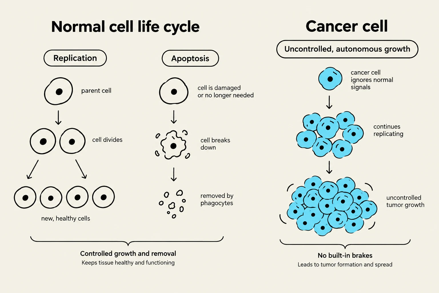
To understand cancer, we must first understand how healthy cells behave. In a normal body, cell replication is a highly regulated process where cells divide to replace old or damaged ones. This is balanced by apoptosis, or programmed cell death, which ensures that damaged or unnecessary cells are removed to prevent harm.
Cells exist in different states of maturity. Stem cells are undifferentiated cells with the unique ability to self-renew and give rise to various cell types. As they begin to specialize, they become progenitor cells, which are more committed to a specific lineage but still capable of division. Eventually, they become differentiated (specialized) cells, which have reached their final functional form (such as a muscle cell or a neuron) and typically lose the ability to divide.
In cancer, this balance is destroyed. The cells exhibit autonomy, meaning they no longer respond to the body's signals to stop growing or to undergo apoptosis. Furthermore, they often exhibit anaplasia, a state where the cells lose their specialized characteristics and appear "undifferentiated" or primitive under a microscope.
This illustration compares the normal cell life cycle, involving replication and apoptosis, with the uncontrolled growth of cancer cells, which leads to tumor formation.
Exam Tip: Remember the distinction between a stem cell (can become anything) and a progenitor cell (already on a specific path toward specialization).
The Genetic Drivers of Malignancy
Cancer is fundamentally a disease of the genome. It is driven by DNA mutations, which can occur during DNA replication, through exposure to environmental damage, or via errors in DNA repair. While some changes are structural, epigenetics refers to changes in gene expression that do not alter the DNA sequence itself but change how the cell "reads" the instructions.
Four specific types of genes play critical roles in this process:
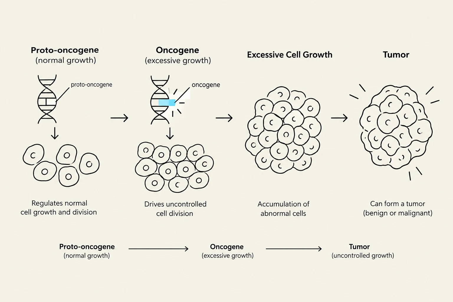
Proto-oncogenes: These are normal genes that help cells grow. When they mutate, they become oncogenes, which act like a "stuck gas pedal," causing continuous cell division.
Tumor Suppressor Genes: These act like "brakes" to slow down cell division. A famous example is the p53 protein. When p53 detects DNA damage, it halts the cell cycle to allow for repair; if the damage is irreparable, p53 triggers apoptosis. If these genes are mutated, the "brakes" fail.
Mutator Genes: These are genes responsible for repairing DNA. If they are damaged, the rate of mutation across the entire genome increases significantly.
This diagram illustrates the progression from a proto-oncogene, which regulates normal cell growth, to an oncogene, which drives uncontrolled cell division, leading to the accumulation of abnormal cells and potentially forming a tumor.
From Initiation to Metastasis
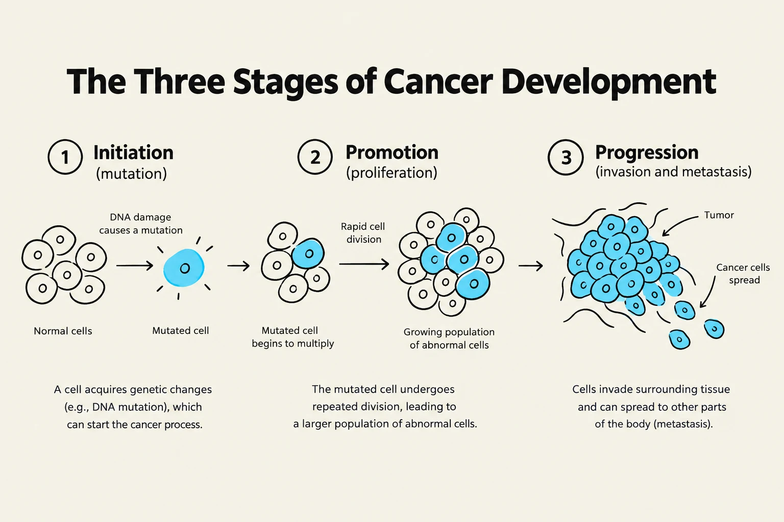
The development of a neoplasm (a new growth or tumor) typically follows three proposed stages:
Initiation: A single cell undergoes a genetic mutation caused by carcinogens (such as chemicals, radiation, or viruses).
Promotion: The mutated cell is stimulated to divide repeatedly, creating a population of abnormal cells.
Progression: The cells continue to mutate and evolve, becoming increasingly aggressive and gaining the ability to invade other tissues.
A neoplasm can be classified as benign (localized, slow-growing, and generally not life-threatening) or malignant (cancerous, invasive, and capable of spreading). Malignant cells spread through several mechanisms: direct extension into adjacent tissues, seeding within a body cavity, or metastasis, where cells travel through the blood or lymph to distant sites. When cancer cells show a preference for a specific organ (e.g., certain cancers frequently spreading to the liver), this is known as organ tropism.
This diagram illustrates the three stages of cancer development: initiation (mutation), promotion (proliferation), and progression (invasion and metastasis).
Clinical Diagnosis and Staging
When a patient presents with a palpable mass, clinicians must determine if it is malignant. This is done through a biopsy (removing tissue for examination) and cytology (the study of individual cells). Doctors may also look for a tumor marker, which is a substance found in blood or urine that is produced by cancer cells.
To standardize how we describe cancer, the TNM classification system is used:
T (Tumor): The size and extent of the primary tumor.
N (Nodes): Whether the cancer has reached nearby lymph nodes (presence of lymphadenopathy or swollen nodes is a key indicator).
M (Metastasis): Whether the cancer has spread to distant parts of the body.
Higher TNM stages generally indicate more advanced disease and a poorer prognosis. Clinicians often track the 5-year survival rate, which includes three groups: patients who are alive, patients who are alive but have had a recurrence, and patients who have died from the cancer.
Treatment and Care
Cancer treatment varies depending on the type and stage, ranging from surgery and radiation to chemotherapy. However, as the disease progresses, patients may experience cachexia, a severe wasting syndrome characterized by weight loss and muscle atrophy.
In advanced stages, the focus often shifts to palliative care. Unlike curative treatments, the goals of palliative care are:
To relieve symptoms and manage pain.
To improve the quality of life for both the patient and their family.
Note on Terminology: In exams, remember that benign tumors often end in "-oma" (e.g., lipoma for fat, adenoma for glandular tissue), whereas malignant tumors often use "-carcinoma" (epithelial origin) or "-sarcoma" (connective tissue origin).
Keep reading for free.
Create an account to finish this reading and make it part of your study routine.