An overview of cancer development, including stages of malignancy, metastasis, and the TNM staging system. It also covers fundamental hematology concepts like anemia and leukemia.
Understanding the progression of cancer and the complexities of hematology is fundamental to clinical practice. Cancer is not a single disease but a process where normal body cells undergo profound genetic and structural changes. This article explores the multi-stage development of malignancy, the physiological mechanisms of metastasis, and the hematological implications of blood disorders such as anemia and leukemia.
Benign vs. Malignant: The Critical Distinction
In oncology, the first step in understanding a tumor is determining its nature. While both involve abnormal cell growth, they differ significantly in their behavior and potential for harm:
Benign Tumors: These are non-cancerous growths. They typically remain localized at their site of origin and do not invade surrounding tissues.
Malignant Tumors: These are cancerous. They possess the ability to invade neighboring tissues and spread to distant parts of the body.
Clinical Note: A key distinction for students is that while "carcinoma in situ" (CIS) involves abnormal cells that have not yet spread, it is considered preinvasive and may eventually become invasive cancer.
The Four Stages of Cancer Development
The transformation from a healthy cell to a life-threatening malignancy follows a predictable, multi-stage progression:
Initiation: The starting point where normal cells undergo genetic mutations or changes.
Promotion: The stage where these initiated cells receive stimulation to begin proliferating.
Progression: The phase where the tumor becomes increasingly advanced, aggressive, and complex.
Metastasis: The final and most dangerous stage, where cancer cells migrate to distant sites.
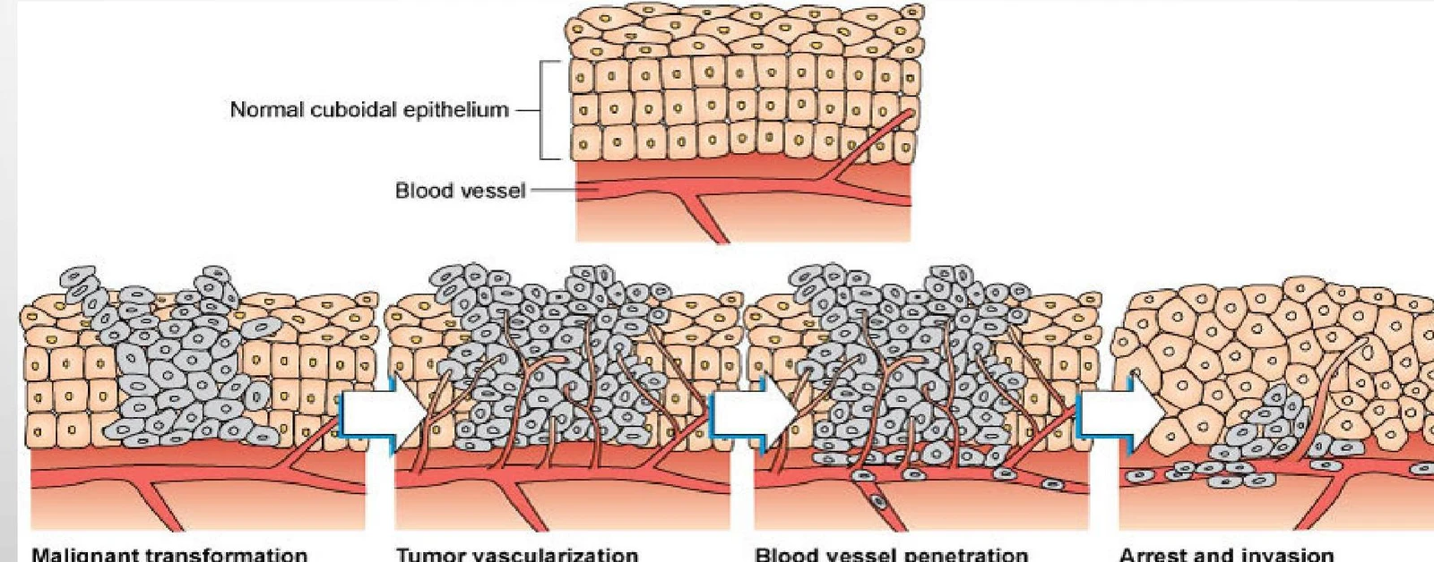
Diagram illustrating the stages of cancer progression, including malignant transformation, tumor vascularization, blood vessel penetration, and arrest and invasion.
The Physiological Path to Metastasis
Metastasis is not a single event but a complex series of physiological maneuvers that allow cancer to move through the body.
Malignant Transformation and Vascularization
The process begins with malignant transformation, where normal cells (such as cuboidal cells in an epithelium) divide excessively to form a tumorous mass. As the tumor grows, it requires its own blood supply to survive. To achieve this, cancer cells secrete Tumor Angiogenesis Factor (TAF). This factor stimulates existing blood vessels to "bud" and grow new channels directly into the tumor, providing the oxygen and nutrients necessary for continued expansion.
Penetration, Arrest, and Invasion
Once the tumor is vascularized, the cells prepare to leave the primary site. Through blood vessel penetration, cancer cells break away from the main tumor mass. They utilize specialized enzymes on their surfaces to create physical openings in the vessel walls, allowing them to enter the circulatory system.
Once in the bloodstream, the cells travel until they undergo arrest and invasion. During this stage, cancer cells clump within the walls of blood vessels in distant organs. If the new environment provides the necessary conditions to support growth, metastatic tumors will form at these new sites.
Staging and Clinical Application: The TNM System
To guide treatment, clinicians use the TNM Classification system to stage cancer:
T (Primary Tumor): Evaluates the size and extent of the primary tumor (ranging from T0—no evidence, to T1−T4 based on size/extent).
N (Regional Lymph Nodes): Indicates whether the cancer has reached nearby lymph nodes (N0 for none, N1−N3 for varying levels of involvement).
M (Distant Metastasis): Determines if the cancer has spread to distant organs (M0 for no spread, M1 for present).
Worked Example: If a patient is staged as T1,N2,Mx, it means there is a small primary tumor (T1), significant involvement of regional lymph nodes (N2), but distant metastasis cannot be evaluated (Mx).
Hematology: Blood Disorders and Cellular Function
Hematology involves the study of blood components: Red Blood Cells (RBCs), White Blood Cells (WBCs), and Thrombocytes (platelets).
Anemia and Sickle Cell Disease
Anemia is not a primary disease but a condition resulting from various etiologies, such as a lack of RBC production, the destruction of existing RBCs, or an overall deficiency in RBC count.
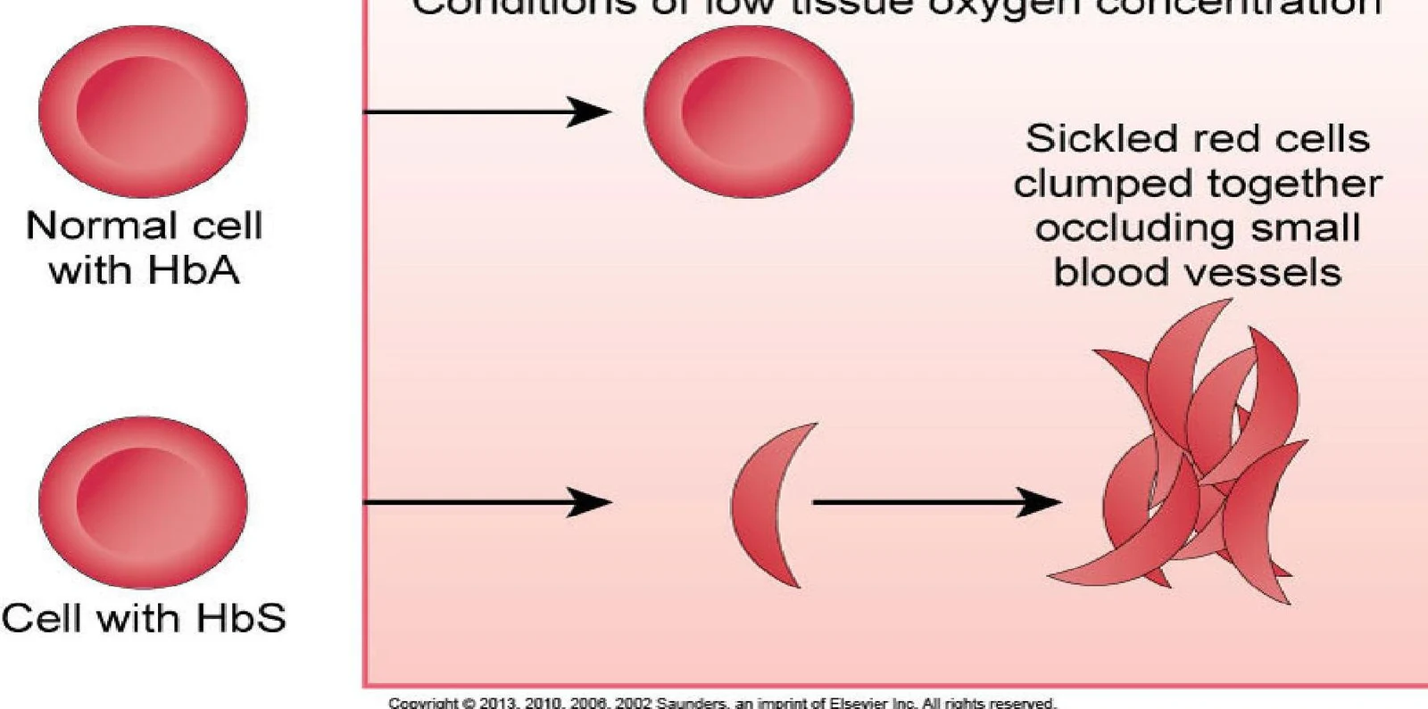
A classic example is Sickle Cell Anemia, an inherited disorder where RBCs become misshapen. During a vasoocclusive crisis, these sickled cells clump together, obstructing blood vessels. This can lead to severe pain, organ infarction (such as in the spleen or kidneys), and hypoxia.
This illustration depicts the difference between normal red blood cells with HbA and sickle-shaped red blood cells with HbS, showing how the latter can clump together and obstruct blood vessels under low oxygen conditions.
Leukemia and White Blood Cell Dynamics
Leukemia is characterized by the uncontrolled production of immature white blood cells in the bone marrow. While the exact cause is often unknown, factors such as ionizing radiation, chemicals, and genetic predispositions play significant roles.
Understanding the WBC count is vital for assessing immune function. The WBCs are often categorized into two "teams":
PMNs (Polymorphonuclear Leukocytes): Including neutrophils, eosinophils, and basophils.
MCS: Including monocytes and lymphocytes.
Exam Tip: When a patient presents with neutropenia (low neutrophils) or thrombocytopenia (low platelets), they are at a significantly increased risk for infection and bleeding, respectively.
Keep reading for free.
Create an account to finish this reading and make it part of your study routine.