Calcium Homeostasis and Endocrine System Disorders
An overview of calcium homeostasis, including the roles of parathyroid hormone, calcitonin, and vitamin D. It also covers endocrine disorders related to the thyroid and adrenal glands, such as hyperparathyroidism, hyperthyroidism, hypothyroidism, Cushing's syndrome, and Addison's disease.
The endocrine system is a sophisticated communication network that maintains internal stability, or homeostasis, through the secretion of hormones. These chemical messengers travel through the bloodstream to target specific cells, triggering physiological responses that can range from rapid metabolic shifts to long-term changes in gene expression. Among the most critical homeostatic processes is the regulation of blood calcium (Ca2+) levels, which are vital for nerve conduction, muscle contraction, and bone integrity. This article explores the intricate feedback loops governing calcium, as well as the clinical manifestations that arise when the thyroid, adrenal, and parathyroid systems malfunction.
Calcium and Bone Homeostasis
Calcium homeostasis is primarily regulated through a tightly controlled feedback loop involving the parathyroid glands, the thyroid gland, and the activation of Vitamin D. Because calcium is essential for various cellular functions, the body prioritizes maintaining a narrow range of concentration in the extracellular fluid.
Parathyroid Hormone (PTH)
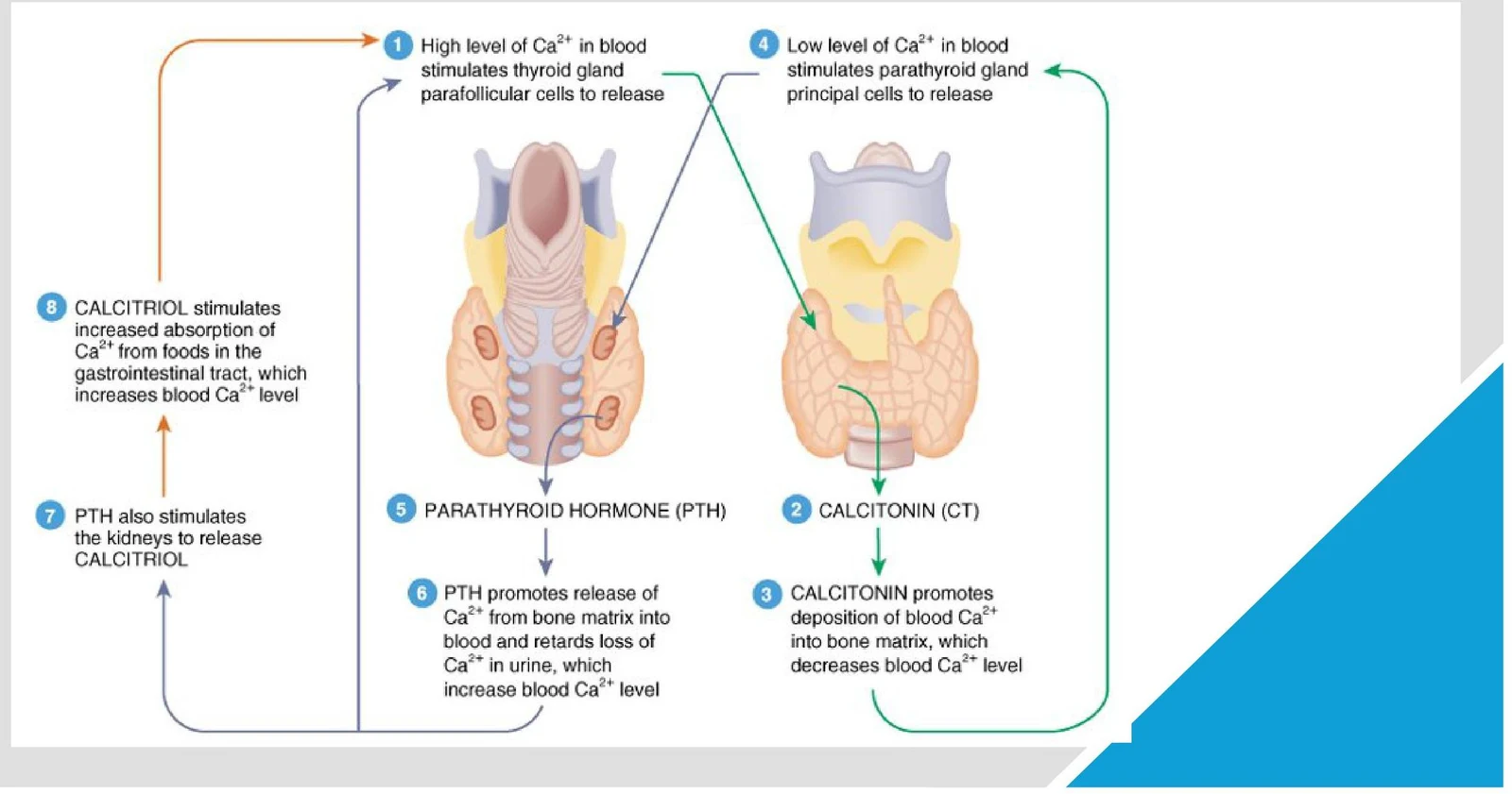
When blood Ca2+ levels drop below the physiological set point, the principal cells of the parathyroid glands detect this change via humoral stimuli. In response, they release Parathyroid Hormone (PTH). PTH acts to increase blood calcium through several mechanisms:
Bone Resorption: PTH hyperactivates osteoclasts, the cells responsible for breaking down bone tissue. This process releases stored calcium from the bone matrix into the bloodstream.
Kidney Function: While not detailed extensively in the summary, PTH also influences the kidneys to assist in calcium retention and the activation of Vitamin D.
Calcitonin (CT)
Conversely, when blood Ca2+ levels are high, the parafollicular cells of the thyroid gland release Calcitonin. Calcitonin acts as an antagonist to PTH, working to lower calcium concentrations and prevent hypercalcemia.
Vitamin D Activation and Calcitriol
Vitamin D is not biologically active immediately upon ingestion or synthesis. It must undergo a series of structural changes, first in the liver and subsequently in the kidneys, to be converted into its active form, Calcitriol. Calcitriol plays a crucial role in increasing blood Ca2+ levels by stimulating the increased absorption of calcium from the gastrointestinal (GI) tract during digestion.
This diagram illustrates the regulation of blood calcium levels by parathyroid hormone (PTH) and calcitonin (CT). High calcium levels stimulate calcitonin release, which lowers blood calcium. Low calcium levels stimulate PTH release, which raises blood calcium. Calcitriol, a hormone derived from vitamin D, also plays a role in calcium absorption.
Exam Tip: The Calcium Feedback Loop
Remember that PTH and Calcitonin are antagonistic. PTH responds to low calcium (to raise it), while Calcitonin responds to high calcium (to lower it).
Hyperparathyroidism
When the parathyroid glands become overactive, they secrete excessive amounts of PTH, a condition known as hyperparathyroidism. This leads to excessive bone resorption, stripping the skeleton of its mineral density. Clinicians often use a specific mnemonic to remember the symptoms of hypercalcemia caused by this disorder: "stones, bones, groans, and psychiatric overtones".
Stones: Kidney stones resulting from high calcium levels in the urine.
Bones: Osteoporosis and increased fracture risk due to bone loss.
Groans: Abdominal pain and gastrointestinal issues.
Psychiatric overtones: Depression, confusion, or lethargy.
A critical edge case is secondary hyperparathyroidism. This often occurs in patients with chronic kidney disease. Because the kidneys are responsible for the final activation of Vitamin D into calcitriol, kidney failure leads to a deficiency in active Vitamin D. The body compensates for the resulting low calcium by overproducing PTH, creating a secondary state of hyperparathyroidism.
Thyroid Regulation and Metabolic Rate
The thyroid gland is a central player in regulating the body's Basal Metabolic Rate (BMR). Its function is governed by the Hypothalamic-Pituitary-Thyroid (HPT) axis: the hypothalamus releases Thyrotropin-Releasing Hormone (TRH), which stimulates the anterior pituitary to release Thyroid-Stimulating Hormone (TSH), which in turn signals the thyroid to produce thyroxine (T4) and triiodothyronine (T3).
Thyrotoxicosis (Hyperthyroidism)
Hyperthyroidism occurs when the thyroid gland is overactive, accelerating the body's metabolic processes. This "speeding up" of metabolism manifests through several clinical signs:
Cardiovascular: Tachycardia (rapid heart rate).
Metabolic: Weight loss and heat intolerance.
Neurological: Tremors and anxiety.
A specific form of hyperthyroidism is Graves' disease, an autoimmune condition where antibodies target the thyroid. A hallmark clinical sign of Graves' disease is exophthalmos, which is the protrusion or bulging of the eyes.
Myxedema (Hypothyroidism)
In contrast, hypothyroidism represents a severe metabolic slowdown. When thyroid hormone levels are insufficient, the body's "engine" slows down, leading to:
Cardiovascular: Bradycardia (slow heart rate).
Metabolic: Weight gain and cold intolerance.
Neurological: Fatigue and mental sluggishness.
This illustration compares the effects of parathyroid and thyroid gland issues, detailing hormones involved, associated symptoms, and common treatments.
Adrenal Function and Stress Response
The adrenal glands, located atop the kidneys, are essential for managing stress and maintaining electrolyte balance. The adrenal cortex is divided into distinct zones that produce different steroid hormones.
Cushing's Syndrome
Cushing's syndrome is caused by chronic excess of cortisol, a glucocorticoid produced in the adrenal cortex. Cortisol is vital for metabolism and the stress response, but in excess, it causes profound systemic changes. Clinical manifestations include:
Central obesity: Fat redistribution to the trunk.
"Buffalo hump": A fatty deposit between the shoulders.
"Moon face": A rounded, swollen facial appearance.
Skin changes: Thinned skin and the appearance of purple stretch marks (striae).
Addison's Disease
Addison's disease is a form of adrenal insufficiency, specifically involving a deficiency in mineralocorticoids like aldosterone. Aldosterone is critical for regulating blood pressure by promoting the retention of sodium and water in the kidneys. When aldosterone is lacking, patients experience:
Profound hypotension: Low blood pressure due to salt and water loss.
Hyperkalemia: A life-threatening buildup of potassium in the blood.
Hyperpigmentation: Darkening of the skin, often due to the co-activation of melanocyte-stimulating hormone.
This illustration depicts various physical symptoms associated with Cushing's syndrome, including moon face, enlarged supraclavicular fat pads, proximal muscular atrophy and weakness, osteoporosis, hypertension, poor wound healing, obesity, abdominal striae, and amenorrhea in women.
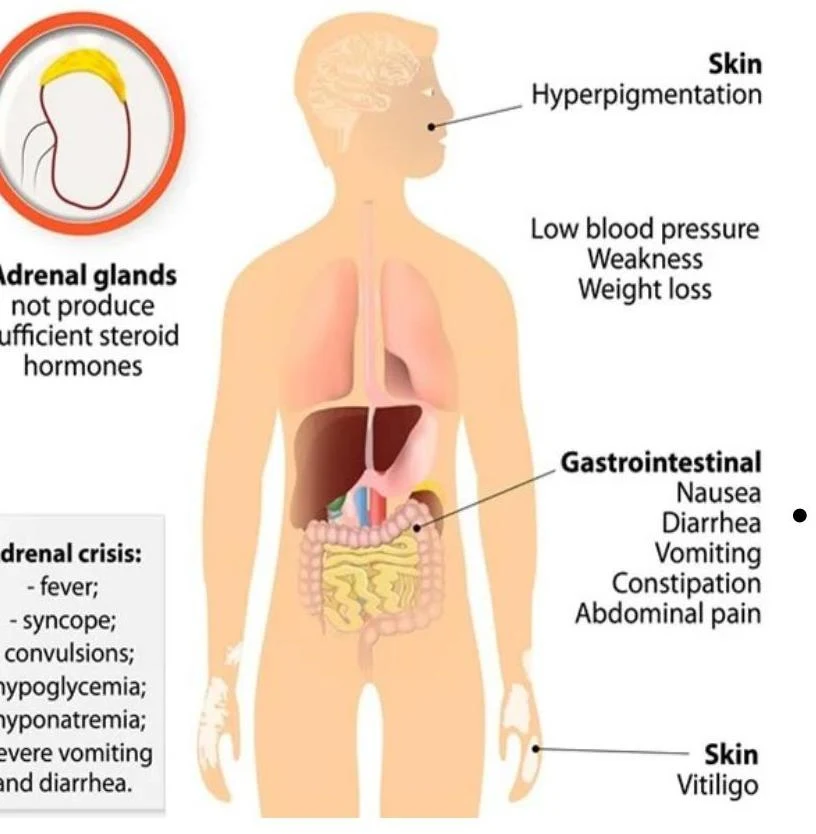
This diagram illustrates the symptoms associated with adrenal insufficiency, including skin changes like hyperpigmentation and vitiligo, gastrointestinal issues, and general symptoms such as low blood pressure, weakness, and weight loss. It also lists symptoms of an adrenal crisis.
Student Pitfall: Primary vs. Secondary Disorders
When diagnosing endocrine issues, always look at the feedback loop. In a primary disorder, the problem is in the target gland (e.g., the thyroid). In a secondary disorder, the problem is in the signaling gland (e.g., the pituitary). For example, in primary hypothyroidism, TSH will be high because the pituitary is trying desperately to "wake up" a failing thyroid. In secondary hypothyroidism, TSH will be low because the pituitary is the source of the failure.
Summary of Hormonal Mechanisms
To understand these disorders, one must understand how hormones work at a cellular level. Hormones are classified by their solubility, which dictates their mechanism of action:
Water-Soluble (Hydrophilic) Hormones: These include peptide and protein hormones (like insulin or TSH). They cannot cross the cell membrane, so they bind to extracellular receptors. This triggers a "second messenger" system (like cAMP) inside the cell to amplify the signal.
Lipid-Soluble (Lipophilic) Hormones: These include steroid hormones (like cortisol, estrogen, and testosterone) and thyroid hormones (T3/T4). They diffuse directly through the cell membrane and bind to intracellular receptors in the cytoplasm or nucleus. This complex then binds to DNA to directly alter gene expression.
By mastering these pathways and the feedback loops that govern them, one can predict how a deficiency or excess in a single hormone will ripple through the entire physiological system.
Keep reading for free.
Create an account to finish this reading and make it part of your study routine.