


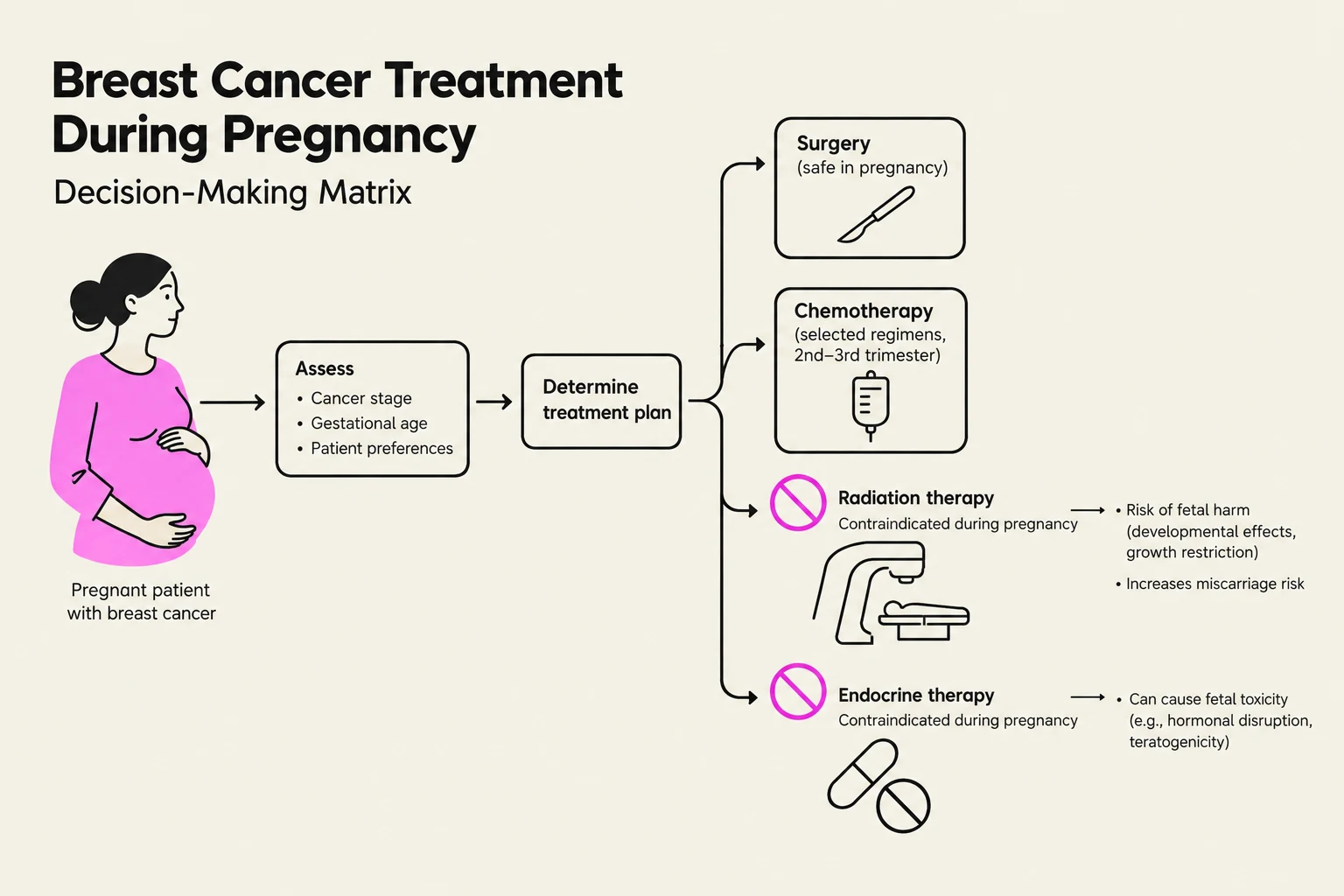
Breast cancer management is a highly individualized process that depends on the specific clinical presentation, the patient's physiological state, and the biological characteristics of the tumor. While standardized staging systems provide a universal language for clinicians, certain subtypes—such as inflammatory breast cancer or Paget disease—and specific patient populations, such as pregnant women, require specialized protocols that deviate from standard surgical or systemic therapy approaches.
Histologic Grading: The Nottingham System
Before determining the stage, pathologists assign a histologic grade to invasive cancers to assess how aggressive the cells appear under a microscope. The most common method is the Nottingham histologic grade, which is a modification of the Scarff-Bloom-Richardson (SBR) system. This grade is calculated based on three key components:
- Tubule formation: How much of the tumor resembles normal breast architecture.
- Nuclear pleomorphism: The variation in size and shape of the nuclei.
- Mitotic count: The rate of cell division.
The resulting score determines the grade:
- G1 (Low grade): Score 3–5; favorable prognosis.
- G2 (Intermediate grade): Score 6–7; moderately favorable.
- G3 (High grade): Score 8–9; unfavorable prognosis.
AJCC TNM Staging System
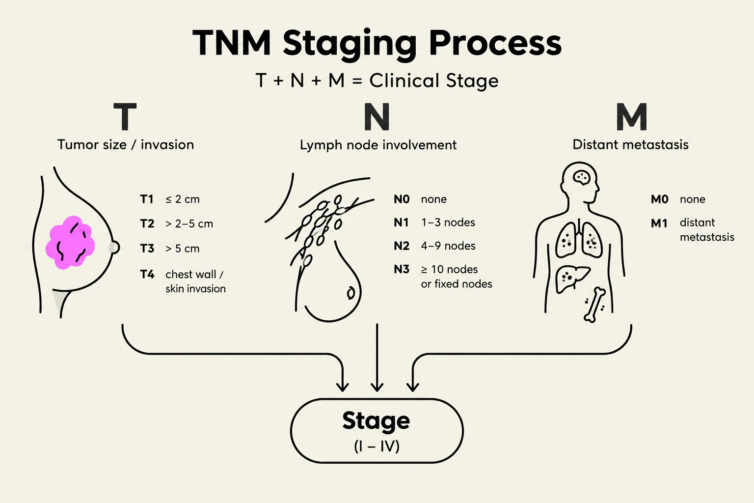
The American Joint Committee on Cancer (AJCC) uses the TNM system to categorize the extent of the disease. This system is essential for determining the stage and selecting appropriate treatment modalities.
T (Primary Tumor)
The T category describes the size and extent of the primary tumor. The most critical distinction is between invasive cancer and carcinoma in situ.
- Tis: Denotes carcinoma in situ, where the cancer is confined to the ducts or lobules and has not invaded the surrounding stroma. This includes Paget disease of the nipple when it is not associated with an underlying invasive cancer.
- T1–T4: These categories are based on the size of the invasive component and whether the tumor has invaded the skin or chest wall.
Keep reading for free.
Create an account to finish this reading and make it part of your study routine.
Continue reading freeAlready have an account? Log in
Updated