Brain Lateralization and Neuropsychological Evidence
Explores brain lateralization, hemispheric specialization, and the role of neuropsychology in understanding brain function through lesion studies and neuroimaging techniques.
How does a single organ, roughly the size of a small melon, manage to coordinate everything from the rhythm of your heartbeat to the complex nuances of a spoken sentence? The human brain is a densely packed, incredibly complex structure containing an estimated 86 billion nerve cells. While the brain functions as a unified whole, research into brain damage and specialized structures reveals that different regions are responsible for distinct cognitive tasks. This concept, known as localization of function, is a cornerstone of modern cognitive psychology.
Lateralization and the Split-Brain Phenomenon
The brain is divided into two roughly symmetrical halves called cerebral hemispheres. While most structures, such as the hippocampus, appear bilaterally (meaning there is one in each hemisphere), the two sides are not identical in their responsibilities. This functional difference is known as lateralization.
The hemispheres are connected by thick bundles of fibers called commissures, the largest of which is the corpus callosum. These fibers allow the two halves to communicate and pool their specialized capacities to produce a single, integrated mental self.
In some medical cases, such as severe epilepsy, surgeons may sever the corpus callosum to limit the spread of electrical disruptions. This results in a split-brain patient—an individual who possesses both brain halves but has severely limited communication between them.
The Evidence of Specialization
Research with split-brain patients has provided profound evidence for hemispheric specialization. Generally, language capacities are lodged in the left hemisphere, while the right hemisphere is crucial for tasks involving spatial judgment.
Consider a classic experimental setup:
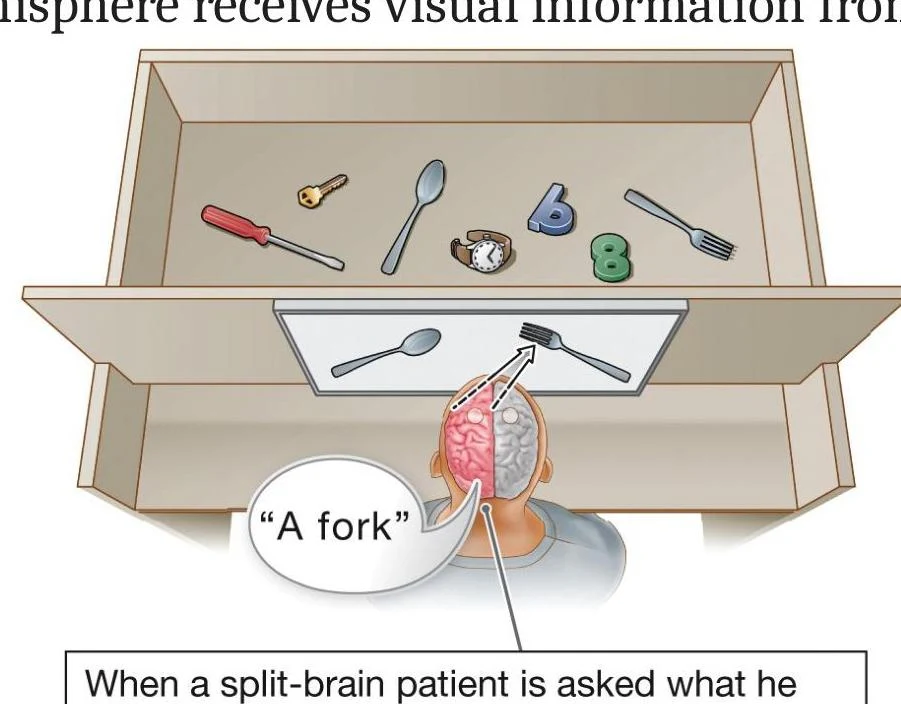
A split-brain patient is shown a fork in their right visual field and a spoon in their left visual field.
If asked to verbally identify the object, the patient will say "fork." This is because the right visual field is processed by the left hemisphere, which controls verbal responses.
However, if asked to pick up the object using their left hand, the patient may pick up the spoon. This occurs because the left hand is controlled by the right hemisphere, which received the visual information from the left side of the world.
Diagram illustrating how visual information is processed in a split-brain patient, showing the left hemisphere receiving input from the right visual field and verbalizing "A fork" when presented with an object in that field.
Illustration depicting how the right hemisphere of the brain processes visual information, specifically showing the perception of a spoon.
Exam Tip: Do not fall for the "left-brain vs. right-brain" myth. While specialization exists, the hemispheres are not competitors; they work as partners to produce complex skills like creativity and intuition.
Clinical Neuropsychology and Lesion Studies
To understand how specific brain structures relate to function, researchers turn to neuropsychology—the study of the brain's structures and their relation to function. A specialized branch, clinical neuropsychology, often utilizes lesion studies. A lesion is a specific area of brain damage that produces observable consequences.
By comparing intact brains to those with damage, scientists can map functions to specific locations. For example, damage to the left side of the frontal lobe typically disrupts language use, whereas damage to the right side rarely has the same effect.
Case Study: Capgras Syndrome
Capgras syndrome (or "imposter syndrome") provides a striking example of how disrupting specific neural circuits can alter reality. Patients with this disorder can recognize their loved ones intellectually but are convinced that the person is an "impostor" or a robot.
This occurs because normal facial recognition requires two separate systems:
Cognitive Appraisal: The ability to identify the person (e.g., "That looks like my father").
Emotional Appraisal: The ability to feel a sense of familiarity (e.g., "That person triggers a warm, safe response").
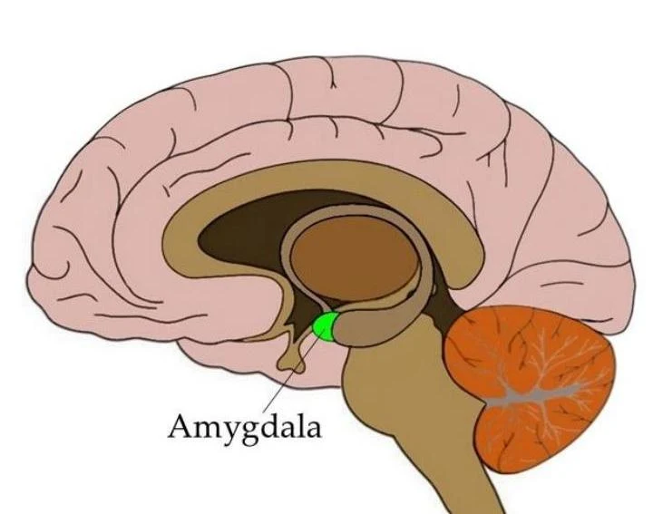
In Capgras syndrome, damage to the right temporal lobe and the amygdala (the brain's "emotional evaluator") disrupts the emotional appraisal. Because the patient feels no emotional "glow" of familiarity, their brain reaches the illogical conclusion that the person must be a fraud. Additionally, damage to the prefrontal cortex can impair the patient's ability to use analytical reasoning to dismiss these bizarre delusions.
Diagram of the amygdala within the human brain.
The Anatomy of the Brain
The brain is organized into three primary structural divisions: the hindbrain, the midbrain, and the forebrain.
The Hindbrain and Midbrain
The hindbrain is located at the top of the spinal cord and manages vital life functions.
Medulla: Controls essential functions like breathing and heart rate.
Pons: Acts as a "bridge" connecting the cerebellum to the rest of the brain.
Cerebellum: Involved in posture, balance, and sensory integration.
The midbrain coordinates precise eye movements, regulates pain, and relays auditory information from the ears to the forebrain.
The Forebrain and Cerebral Cortex
The forebrain is the largest region and contains the cerebral cortex—the thin, wrinkled outer layer of the brain. The cortex is divided into four main lobes:
Frontal Lobe: Responsible for planning, analysis, and decision-making.
Parietal Lobe: Located at the topmost part of the brain.
Temporal Lobe: Involved in auditory processing and memory.
Occipital Lobe: Dedicated to visual processing.
Beneath the cortex lie the subcortical structures, including the thalamus (the sensory relay station), the hypothalamus (which regulates biological needs like hunger), and the limbic system. The limbic system contains the hippocampus, which is essential for forming new memories.
Modern Research: Neuroimaging
While lesion studies provide causal evidence, neuroimaging allows us to observe the living, functioning brain in real-time.
Structural vs. Functional Imaging
Neuroimaging is categorized into two main types:
Structural Imaging: Provides a detailed physical map of the brain's shape and size. Examples include CT scans (3D X-rays) and MRI (which uses magnetic properties of atoms).
Functional Imaging: Tracks brain activity over time.
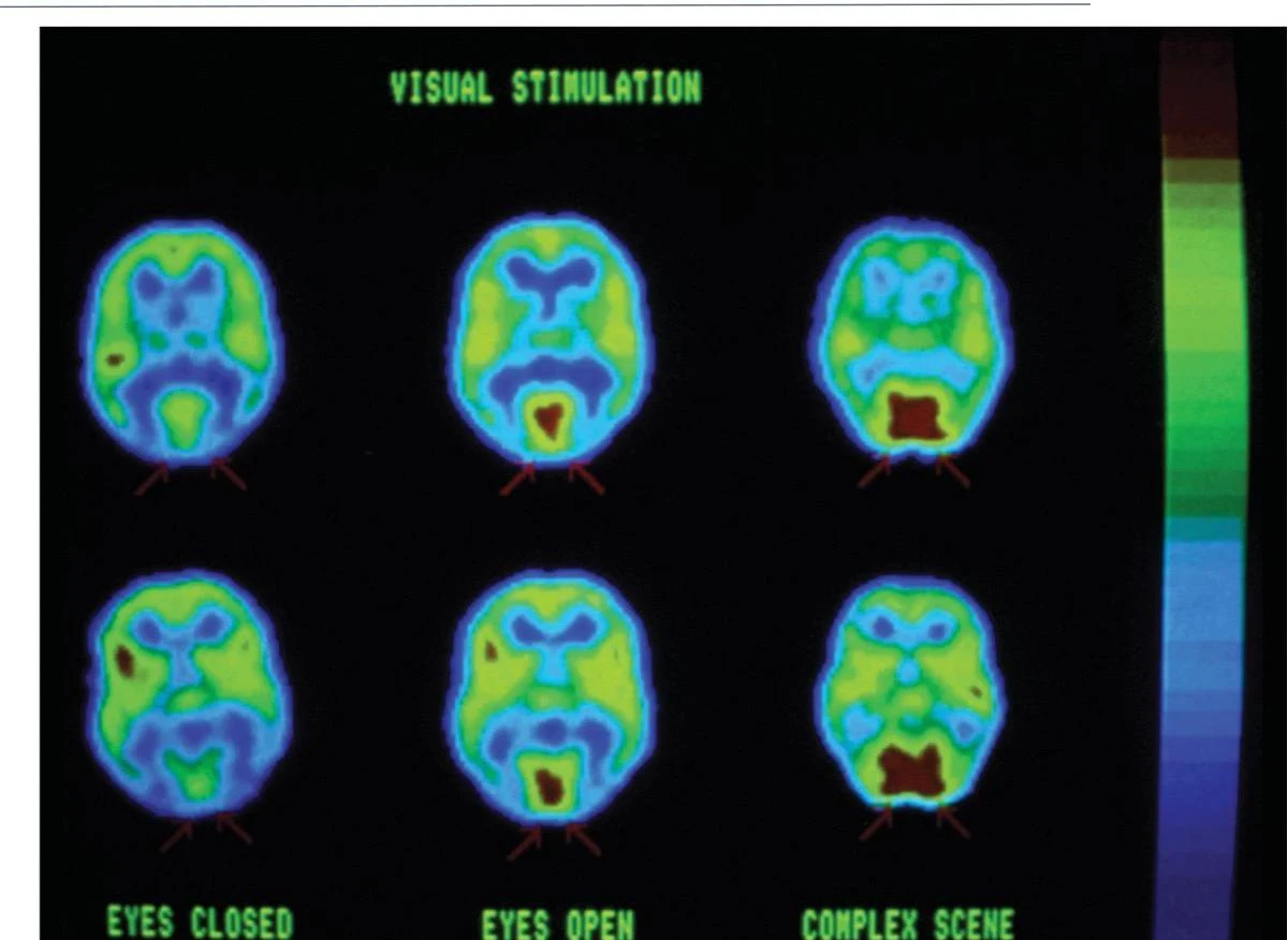
PET Scans: Use a radioactive tracer (like glucose) to show which tissues are most active.
fMRI: Measures changes in oxygen content in the blood to provide a "video clip" of moment-by-moment neural activity.
Brain activity scans under different visual stimulation conditions: eyes closed, eyes open, and viewing a complex scene.
Functional imaging has allowed researchers to identify highly specialized areas, such as the fusiform face area (FFA) for face recognition and the parahippocampal place area (PPA) for recognizing places.
Summary Table: Research Methods
Method
Strength
Weakness
EEG
Excellent temporal resolution (When?)
Poor spatial resolution (Where?)
MRI
Excellent spatial resolution (Where?)
Low temporal resolution (When?)
fMRI
High spatial resolution (Where?)
Moderate temporal resolution (When?)
Keep reading for free.
Create an account to finish this reading and make it part of your study routine.