Brain Function and Domestic Violence: Neurological Basis of Impulse Control
Examines the neurological basis of impulse control and its relation to domestic violence, exploring how brain electrical activity, cellular malfunctions, and genetic factors contribute to rage and loss of self-control. Discusses medical interventions like medication and genetic mapping.
When we witness an episode of domestic violence—a sudden, explosive outburst of rage over a minor provocation—the immediate social reaction is often to label the individual as having a "bad character" or a "flawed personality". However, modern medical science is shifting this perspective. Rather than viewing these episodes solely as moral failings, researchers are increasingly looking at the biological malfunctions within the brain that prevent individuals from regulating their emotions. This article examines the intersection of neurology and behavior, specifically focusing on how electrical irregularities can lead to a total loss of self-control.
The Neurology of Rage: Electrical Activity and Voltage
At the core of emotional regulation is the brain's ability to process information and manage stress through electrical signaling. In a healthy brain, when a person encounters a problem or a social provocation, specific areas of the brain activate to "solve" the issue through communication and cognitive reasoning.
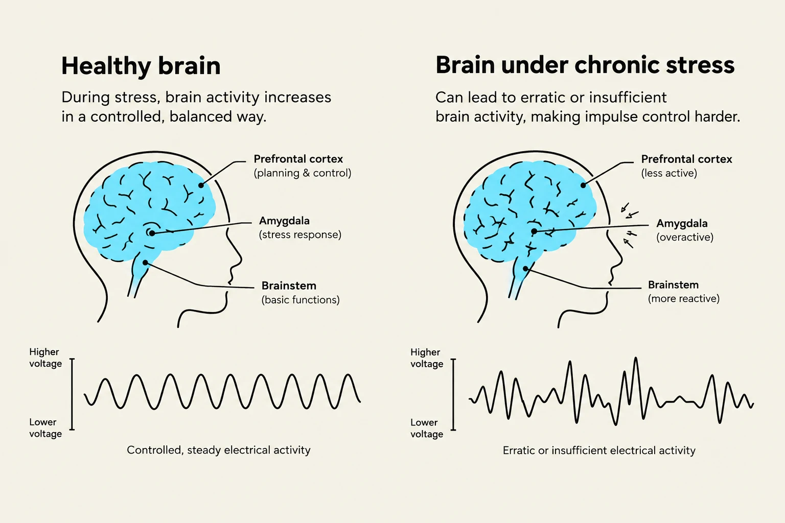
This process is measurable through brain scans, which monitor the voltage levels of electrical activity. A healthy response to stress involves a certain threshold of electrical output that allows the brain to navigate frustration without escalating into violence. In individuals prone to explosive rage, this electrical "budget" is often mismanaged. Some patients may exhibit much higher voltage spikes in response to minor stressors, while others may lack the necessary electrical engagement to process the situation rationally.
Comparison of brain activity in a healthy brain versus a brain under chronic stress, illustrating the impact on the prefrontal cortex, amygdala, and brainstem, along with corresponding electrical activity patterns.
Cellular Malfunction and the "Out of Kilter" Brain
When we look closer at the cellular level, the cause of these outbursts becomes even more apparent. Research into Intermittent Explosive Disorder suggests that the issue is not just about the amount of electricity, but the functionality of the cells themselves.
In some patients, brain scans reveal very little electrical activity in critical regulatory regions. This indicates a cellular malfunction: the cells are not firing or processing information correctly. When these cells fail to communicate, the brain becomes "out of kilter"—it is no longer firing in a synchronized or healthy manner. This lack of synchronization means the brain cannot effectively bridge the gap between a stimulus (like a late appointment or a minor disagreement) and a controlled response, leading to an immediate explosion of rage.
Exam Tip: If asked to explain the difference between a "character flaw" and a "biological malfunction," focus on the distinction between intent and neurological capacity. A character flaw implies a choice, whereas a biological malfunction (like cellular non-firing) implies an inability to process information correctly due to physiological limits.
Medical Interventions: Stabilizing the System
Because these outbursts are linked to biological irregularities, the solutions are increasingly moving toward medical intervention rather than purely disciplinary measures.
Pharmacological Treatment
One of the most significant breakthroughs involves the use of medications to "jump-start" or stabilize the brain's response to stress. Specifically, beta-blockers have shown dramatic results in clinical settings. By managing the physiological and electrical symptoms of rage, these drugs help stabilize the individual, allowing them to regain a level of control that was previously impossible.
Genetic Mapping and Behavior
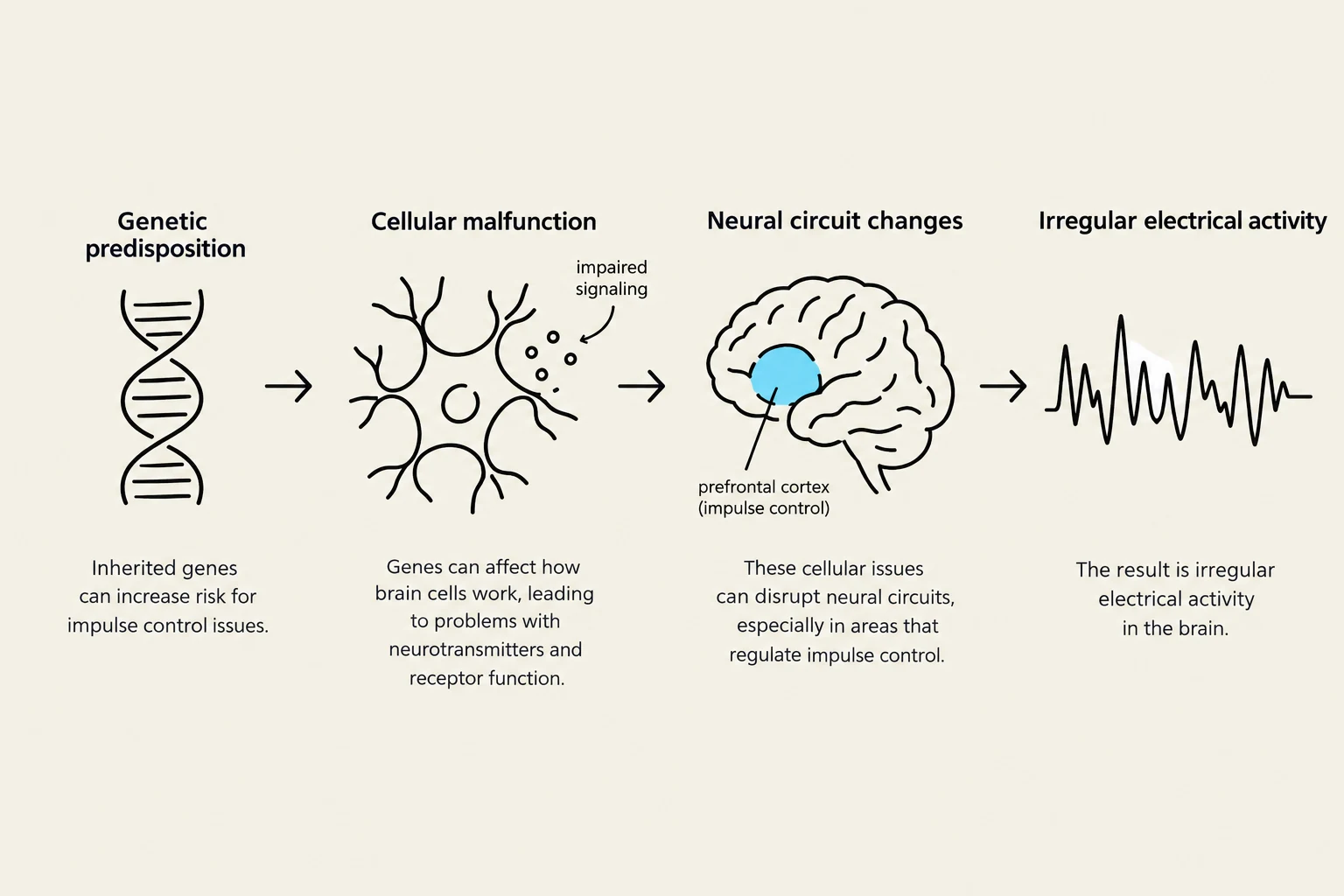
Beyond immediate medication, scientists are working on the long-term identification of these disorders through biological mapping. By mapping out an individual's genetics, researchers hope to understand the hereditary predispositions to these neurological malfunctions. Understanding the intersection of genetics and brain behavior could allow for earlier intervention before domestic violence patterns become established.
This diagram illustrates how genetic predisposition can lead to cellular malfunction, neural circuit changes, and ultimately irregular electrical activity in the brain, potentially affecting impulse control.
Summary of the Biological Theory
The theory presented suggests that domestic violence can be a symptom of a profound biological failure. When the brain fails to provide the necessary electrical regulation to manage frustration, the individual loses the ability to self-regulate.
To summarize the relationship between these variables, we can consider the following conceptual model:
Biological Malfunction→Cellular Non-firing→Electrical Irregularity→Loss of Impulse Control→Domestic Violence
By treating the underlying neurological cause—rather than just the outward behavior—medical science offers a path toward reducing domestic violence and helping families stabilize.
Study Note: When reviewing for the exam, ensure you can link the three levels of dysfunction:
Cellular (cells not firing)
Electrical (voltage/activity issues)
Behavioral (loss of control/violence).
Keep reading for free.
Create an account to finish this reading and make it part of your study routine.