This article explains the distribution of body water, the mechanisms of solute transport, and the clinical application of the anion gap, covering concepts like osmosis, ion gradients, and electrochemical balance.
Understanding how water and electrolytes move between body compartments is fundamental to clinical medicine. Whether you are managing a patient with dehydration, heart failure, or metabolic acidosis, the principles of osmosis, ion gradients, and electrochemical balance dictate the physiological response. This article explores the distribution of body water, the mechanisms of solute transport, and the clinical application of the anion gap.
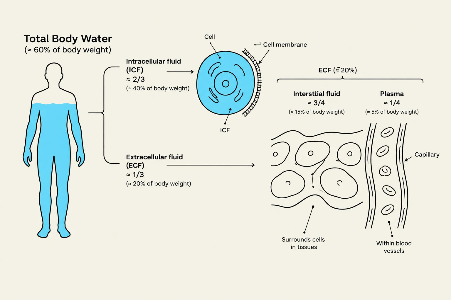
Total Body Water and Fluid Compartments
In a healthy, lean 70-kg adult, Total Body Water (TBW) accounts for approximately 60% of body mass, which equates to about 42 L. However, this percentage is not static; it varies significantly based on age and body composition. Infants possess a higher fraction of body water, whereas aging and increased adiposity (fat content) decrease the total percentage. Clinically, it is useful to remember that a rapid weight change of 1 kg is roughly equivalent to a 1 L change in net fluid volume.
Body fluids are organized into distinct compartments separated by semi-permeable barriers:
Intracellular Fluid (ICF): The fluid contained within the cell, separated from the outside by the cell membrane.
Extracellular Fluid (ECF): The fluid outside the cells, which is further divided into:
Interstitial Fluid: The fluid surrounding the cells.
Plasma: The liquid component of blood, separated from the interstitial fluid by the capillary wall.
Diagram illustrating the distribution of total body water, differentiating between intracellular fluid (ICF) and extracellular fluid (ECF), and further detailing ECF into interstitial fluid and plasma.
Osmosis and Osmolality
The movement of water between these compartments is driven by osmosis. Water moves passively from areas of low effective solute concentration to areas of high effective solute concentration. While water can cross the lipid bilayer of cell membranes at a slow rate, the process is significantly accelerated by specialized protein channels called aquaporins, which increase membrane permeability.
It is vital to distinguish between osmolality (the total concentration of all dissolved particles) and tonicity (the concentration of "effective" or non-penetrating solutes that cause water to move). For example, urea can increase osmolality because it is a solute, but it may not change cell volume significantly because it can equilibrate across the membrane, whereas solutes like sodium cannot.
To estimate plasma osmolality in a clinical setting, the following formula is often used:
Calculated Osmolality≈2×Na++18Glucose+2.8BUN
In this equation, Na+ is in mmol/L, while glucose and BUN are in mg/dL. If the measured osmolality is significantly higher than this calculated value, it indicates the presence of "unmeasured osmoles," such as ethanol or toxic alcohols (an osmolal gap).
Ion Distribution and the Na+/K+ Pump
The distribution of ions is highly specific and maintained by active transport mechanisms. The ECF is characterized by high concentrations of Na+ (135–145 mEq/L) and Cl− (98–107 mEq/L), while the ICF is rich in K+, Mg2+, and proteins.
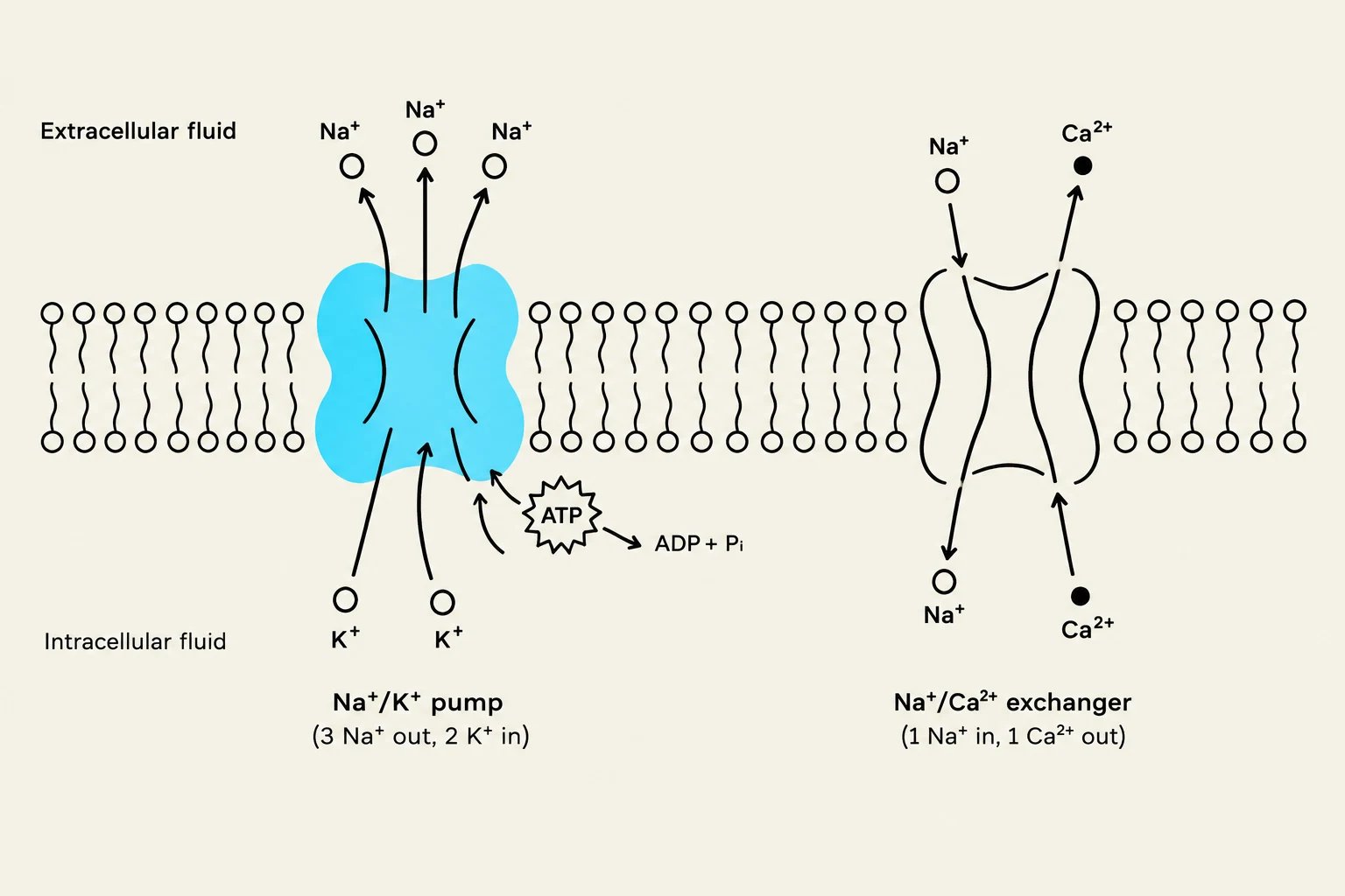
The primary driver of these gradients is the Na+/K+-ATPase pump. This pump uses ATP to perform primary active transport, exporting 3Na+ ions for every 2K+ ions it imports. This process creates two critical conditions:
A low intracellular Na+ concentration and a high intracellular K+ concentration.
A net outward movement of positive charge, contributing to the membrane potential.
These stored gradients provide the energy for secondary active transport. For instance, the Na+/Ca2+ exchanger (NCX) uses the Na+ gradient to export Ca2+ from the cell. This connection is clinically relevant in pharmacology; for example, digoxin inhibits the Na+/K+-ATPase, leading to increased intracellular Na+, which subsequently reduces the efficiency of the Na+/Ca2+ exchanger, ultimately increasing intracellular Ca2+ and boosting cardiac contractility.
Diagrams illustrating the Na+/K+ pump and the Na+/Ca2+ exchanger, both transmembrane protein complexes involved in ion transport across cell membranes.
Plasma Proteins and Oncotic Pressure
Within the plasma, proteins play a crucial role in maintaining fluid balance. Albumin is the most abundant plasma protein and is responsible for providing the majority of plasma oncotic pressure (also known as colloid osmotic pressure). This pressure acts as a "pulling" force that keeps fluid within the vascular compartment.
When albumin levels drop (hypoalbuminemia), the oncotic pressure decreases, which can lead to fluid leaking into the interstitial space, resulting in edema or ascites.
Electroneutrality and the Serum Anion Gap
In any biological solution, the principle of electroneutrality must be maintained: the sum of all positive charges must equal the sum of all negative charges. In the serum, we can calculate the anion gap (AG) to identify "unmeasured" anions that are not part of the standard laboratory panel (Cl− and HCO3−).
The formula is:
AG=Na+−(Cl−+HCO3−)
A typical normal interval is approximately 8–12 mEq/L.
An increased anion gap suggests the accumulation of unmeasured acid anions. Common causes include:
Lactate (e.g., from septic shock).
Ketoanions (e.g., from diabetic ketoacidosis).
Toxic alcohols (e.g., methanol or ethylene glycol).
Changes in Albumin: Since albumin is a major unmeasured anion, a significant drop in albumin will decrease the measured anion gap.
Exam Tip: If a patient presents with a high anion gap and metabolic acidosis, always look for "MUDPILES" (Methanol, Uremia, Diabetic Ketoacidosis, Propylene glycol, Isoniazid, Lactic acidosis, Ethylene glycol, Salicylates) as potential culprits.
Clinical Summary: Fluid Shifts and Adaptation
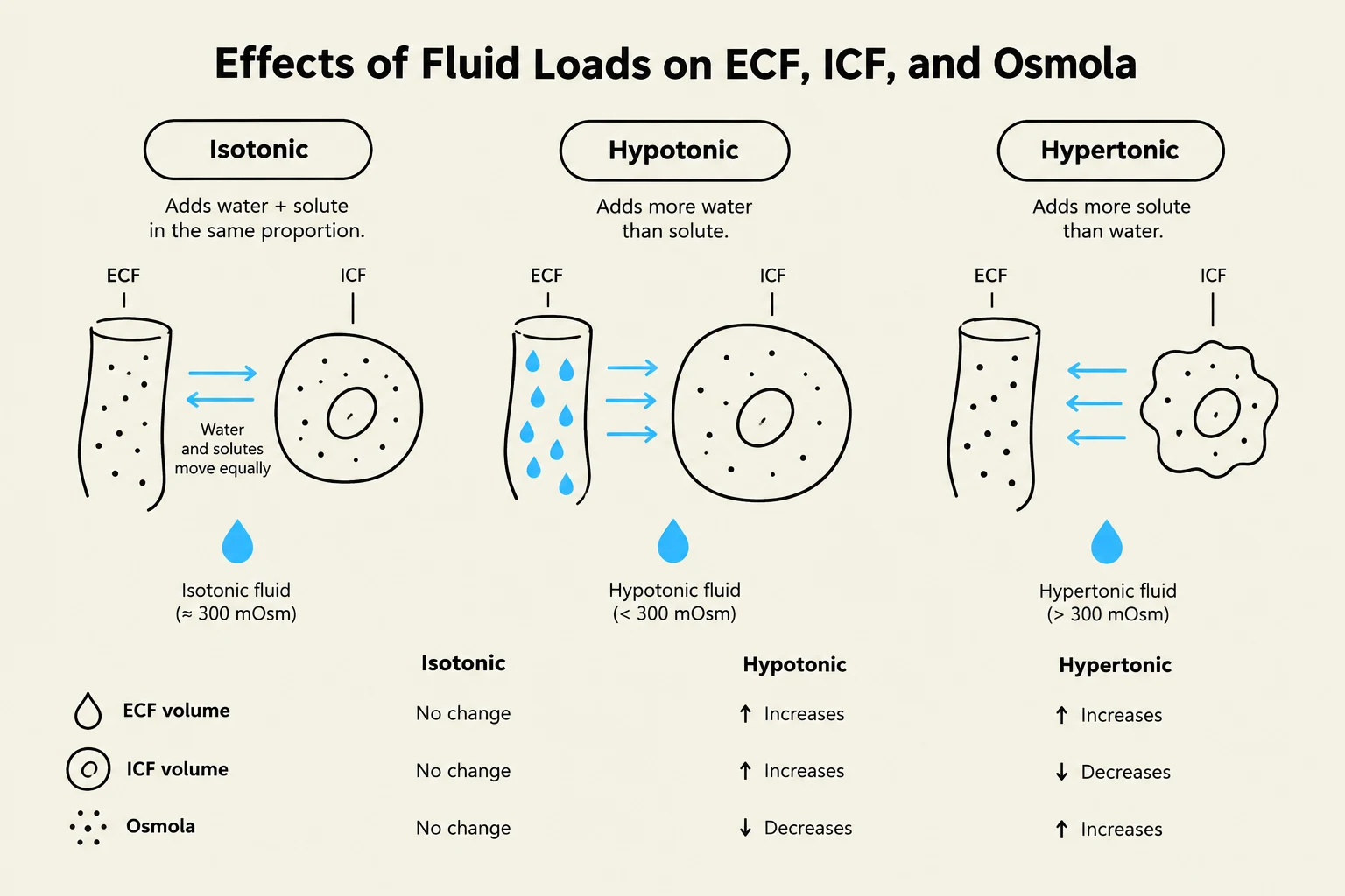
The clinical impact of fluid changes depends on whether the loss or gain is isotonic, hypotonic, or hypertonic.
Fluid Type
Effect on ECF
Effect on ICF
Osmolality
Isotonic (e.g., 0.9% NaCl)
Increases
No change
No change
Hypotonic (e.g., 0.45% NaCl)
Increases
Increases
Decreases
Hypertonic (e.g., 3% NaCl)
Increases
Decreases
Increases
This diagram illustrates the effects of isotonic, hypotonic, and hypertonic fluid loads on extracellular fluid (ECF) and intracellular fluid (ICF) volumes and osmolarity. Isotonic fluids maintain equilibrium, hypotonic fluids cause cells to swell, and hypertonic fluids cause cells to shrink.
The brain is particularly sensitive to these shifts. In acute hyponatremia (low sodium), water rushes into brain cells, causing swelling (cerebral edema), which can lead to seizures or coma. If the body has had time to adapt to chronic hyponatremia, it does so by exporting organic osmolytes to reduce intracellular pressure. However, if such a patient is corrected too rapidly, the brain cells—now lacking those osmolytes—will shrink too quickly, potentially causing osmotic demyelination syndrome.
Keep reading for free.
Create an account to finish this reading and make it part of your study routine.