Blood, Lymph, and Immunity: Quantitative Evaluation and Clinical Terminology
An overview of blood composition, hematology, and the immune system, including quantitative evaluation through Complete Blood Count (CBC) and terminology for blood and immune disorders.
Understanding the composition and function of blood is fundamental to veterinary and medical science. Blood is classified as a connective tissue that serves as a vital transport medium for nutrients, gases, and immune cells. By utilizing quantitative tools like the Complete Blood Count (CBC), clinicians can assess the health of an individual, identify infectious diseases, and monitor the body's immune response.
The Complete Blood Count (CBC) and Red Blood Cell Indices
A Complete Blood Count (CBC) is a comprehensive panel used to evaluate the various cellular components of blood. This includes red blood cells (RBCs), white blood cells (WBCs), and platelets.
Red Blood Cell (RBC) Parameters
To understand the oxygen-carrying capacity of the blood, several specific metrics are analyzed:
RBC Count: The total number of red blood cells per unit of volume.
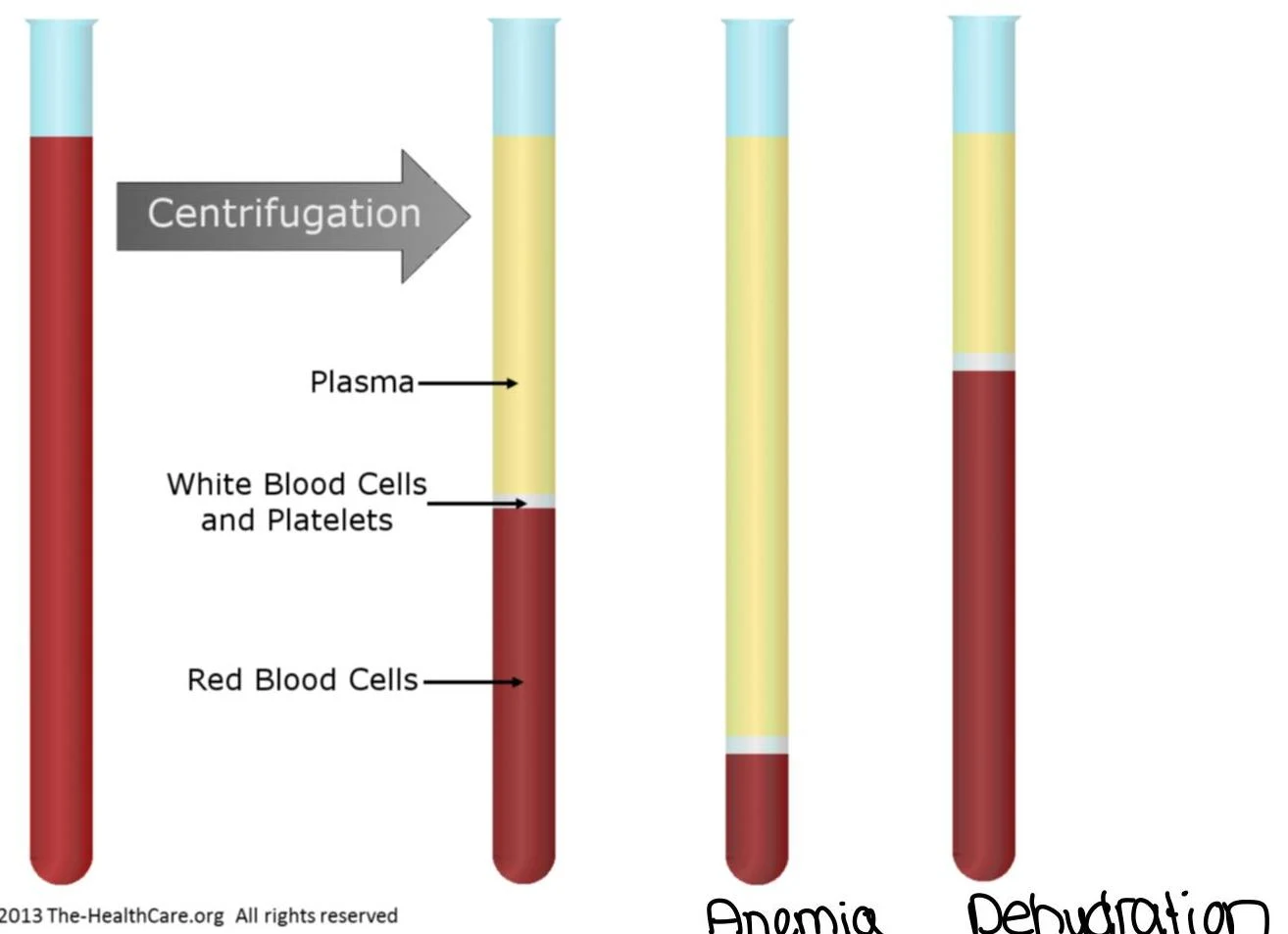
Hematocrit (PCV): Also known as Packed Cell Volume, this measures the percentage of total blood volume occupied by red blood cells.
A hematocrit tube showing the separation of blood components, with red blood cells making up 45% of the total volume.Diagram illustrating blood separation by centrifugation, showing normal blood composition, anemia (reduced red blood cells), and dehydration (reduced plasma).
Reticulocyte Count: This measures immature red blood cells, providing insight into the bone marrow's ability to produce new cells (erythropoiesis).
Red Cell Distribution Width (RDW): This indicates the variation in size among the red blood cells.
To further characterize the quality of the RBCs, clinicians look at "indices":
Mean Corpuscular Volume (MCV): The average size of a single red blood cell.
Mean Corpuscular Hemoglobin (MCH): The average amount of hemoglobin per red blood cell.
Mean Corpuscular Hemoglobin Concentration (MCHC): The average concentration of hemoglobin in a given volume of packed red cells.
Clinical Terminology for RBC Disorders
When these values fall outside the normal range, specific medical terms are used:
Anemia: A deficiency in red blood cells or hemoglobin, often resulting in clinical signs like jaundice or weakness.
Erythrocytosis / Polycythemia: An abnormally high concentration of red blood cells.
Erythroblastopenia: A deficiency in the production of erythroblasts (red blood cell precursors).
White Blood Cell (WBC) Analysis and the Differential Count
The White Blood Cell (WBC) count measures the total number of leukocytes in the blood, which is a primary indicator of infection or inflammation. A WBC differential count breaks this total down into specific types of cells, each with a unique role in the immune system.
The Leukocyte Types
Leukocytes are broadly categorized into granulocytes and agranulocytes:
Granulocytes (contain visible granules):
Neutrophils: The most common WBC, primarily responsible for fighting bacterial infections.
Eosinophils: Often involved in allergic reactions and fighting parasitic infections.
A micrograph shows a neutrophil, a type of white blood cell, surrounded by red blood cells.
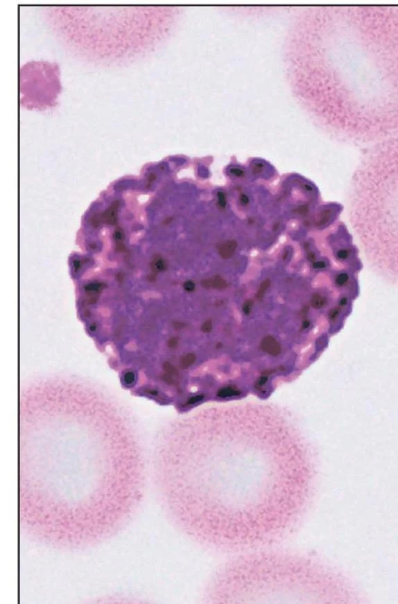
Basophils: Involved in inflammatory responses.
A microscopic view of a basophil, a type of white blood cell, surrounded by red blood cells.
Agranulocytes:
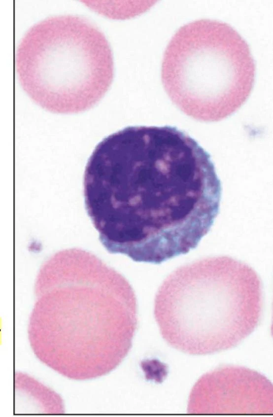
Lymphocytes: Key players in the adaptive immune system, including B cells and T cells.
A microscopic view of a lymphocyte surrounded by red blood cells.
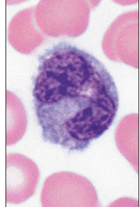
Monocytes: Large cells that can differentiate into macrophages to engulf pathogens.
A microscopic view of a monocyte, a type of white blood cell, surrounded by red blood cells.
Clinical Terminology for WBC Disorders
Leukocytosis: An increase in total WBC count.
Leukopenia: A decrease in total WBC count.
Granulocytosis / Granulocytopenia: An increase or decrease in granulocytes.
Agranulocytosis: A severe deficiency of granulocytes.
Specific Cell Fluctuations:
Neutrophilia / Neutropenia: High or low neutrophil counts.
Lymphocytosis / Lymphocytopenia: High or low lymphocyte counts.
Eosinophilia / Eosinopenia: High or low eosinophil counts.
Basophilia / Basopenia: High or low basophil counts.
Platelets and Hemostasis
Platelets (Thrombocytes) are cell fragments essential for blood clotting (hemostasis).
Thrombocytosis: An abnormally high platelet count.
Thrombocytopenia: An abnormally low platelet count.
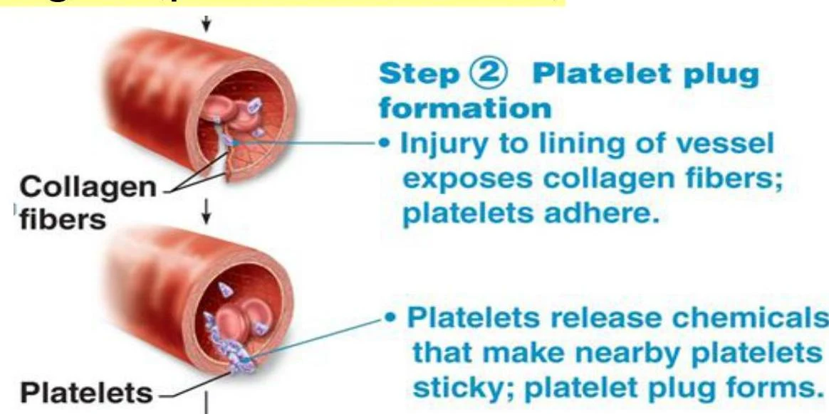
The process of clotting involves a complex cascade where platelets form a plug and fibrin strands create a mesh to trap cells and stabilize the clot:
Diagram illustrating the formation of a platelet plug in response to a blood vessel injury, showing collagen exposure and platelet aggregation.Diagram illustrating the final stages of blood coagulation, showing the conversion of fibrinogen to fibrin and the formation of a cross-linked fibrin mesh.
Note on Pancytopenia: If a patient presents with a simultaneous decrease in RBCs, WBCs, and platelets, the condition is termed pancytopenia.
The Immune System: Humoral vs. Cellular Immunity
The immune system is divided into two main branches of adaptive immunity: Humoral Immunity and Cellular Immunity.
Humoral Immunity (B Cells)
Humoral immunity is mediated by B lymphocytes that differentiate into plasma cells to secrete antibodies (immunoglobulins). These antibodies target extracellular pathogens.
Different classes of antibodies serve different roles:
IgM: The first antibody produced during an initial infection (acute phase).
IgG: The most abundant antibody in serum; high levels often indicate a chronic infection or a secondary immune response.
IgE: Primarily involved in allergic reactions by binding to mast cells.
Cellular Immunity (T Cells)
Cellular immunity is mediated by T lymphocytes, which target intracellular pathogens (like viruses):
Helper T-cells: Release chemicals to activate other lymphocytes or macrophages.
Killer (Cytotoxic) T-cells: Directly destroy infected or cancerous cells.
Memory T-cells: Ensure a faster response upon re-exposure to a pathogen.
Immunological Memory and Vaccination
The primary immune response occurs upon first exposure to an antigen, characterized by a lag period and lower antibody titers. The secondary immune response occurs upon re-exposure; it is much faster and more robust due to the presence of memory cells.
Vaccinations exploit this mechanism by providing "artificially acquired active immunity," stimulating the body to create memory cells without the animal having to suffer the full disease.
Exam Tip: Remember the distinction between Active and Passive immunity.
Active: The body makes its own antibodies (e.g., through infection or vaccine).
Passive: The body receives antibodies from an outside source (e.g., colostrum from a mother or an injection of gamma globulins). Passive immunity provides immediate protection but lacks immunological memory.
Keep reading for free.
Create an account to finish this reading and make it part of your study routine.