Blood Composition and Hemostasis: The Body's Vital Fluid
An overview of blood's composition, including plasma and formed elements (erythrocytes, leukocytes, platelets), and its crucial roles in transport, regulation, and protection within the human body. It also details the process of hemostasis, the mechanism by which bleeding is stopped.
Blood is a unique, specialized connective tissue that exists in a fluid state, serving as the primary medium for internal transport within the human body. It is essential for maintaining life, acting as a bridge between various organ systems to ensure that cells receive what they need and dispose of what they cannot use.
The Three Pillars of Blood Function
Blood performs three fundamental roles: transport, regulation, and protection.
1. Transport
Blood acts as a high-speed delivery and waste removal system.
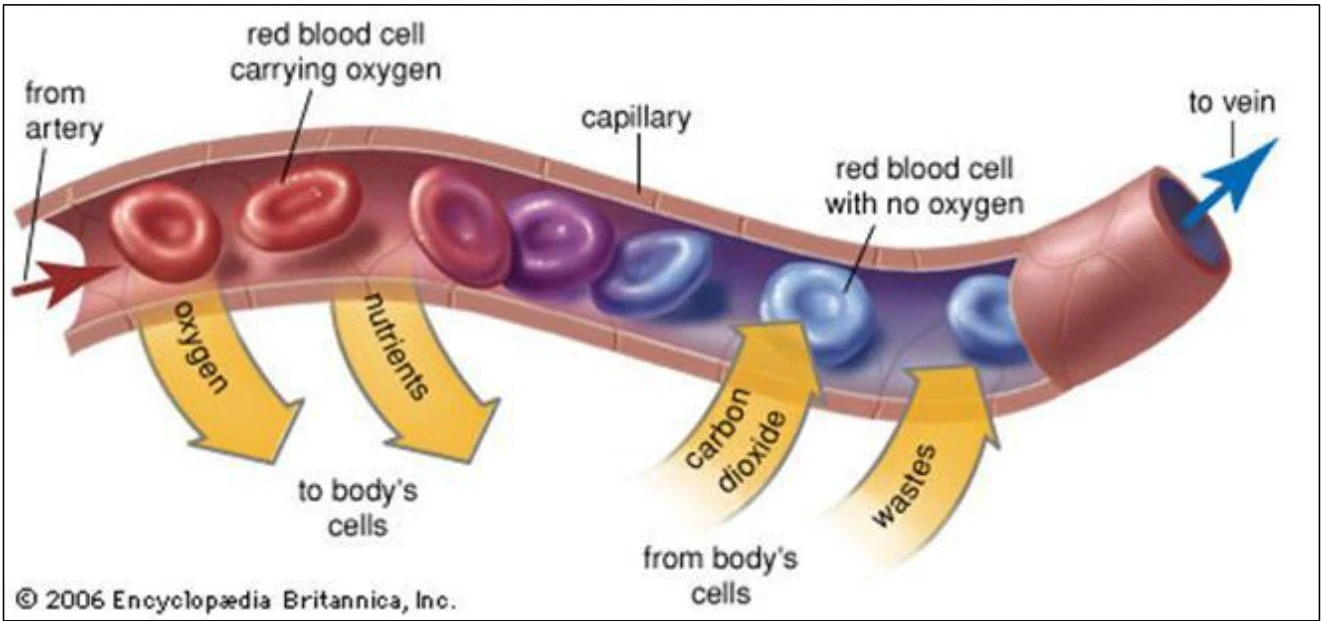
Nutrients and Gases: It delivers oxygen (O2) and nutrients to body cells while simultaneously carrying metabolic wastes to the lungs and kidneys for elimination.
Hormones: It serves as a communication highway, transporting hormones from endocrine organs to their specific target organs.
Diagram illustrating the exchange of oxygen, nutrients, carbon dioxide, and wastes between red blood cells in a capillary and body cells.
2. Regulation
Blood helps maintain a stable internal environment (homeostasis) through several mechanisms:
Temperature: It absorbs and distributes heat throughout the body to maintain a steady temperature.
pH Balance: It uses buffers, such as the alkaline reserve of bicarbonate ions, to maintain normal pH levels.
Fluid Volume: It helps maintain adequate fluid volume within the circulatory system.
3. Protection
The blood protects the body from two major threats: blood loss and infection.
Preventing Blood Loss: Plasma proteins and platelets work together to initiate clot formation when a vessel is injured.
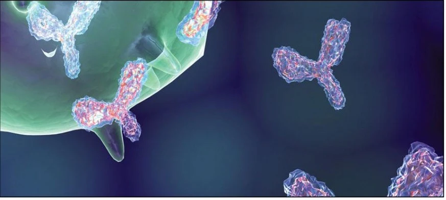
Preventing Infection: The blood carries the body's primary defense agents, including antibodies, complement proteins, and white blood cells.
Antibodies interacting with a cell.
Composition of Blood
Blood is composed of two main components: a non-living fluid matrix called plasma and living cells known as formed elements.
Plasma: The Liquid Matrix
Plasma is a straw-colored, sticky fluid that makes up the bulk of the blood. It is approximately 90% water and contains over 100 dissolved solutes, including electrolytes, nutrients, gases, and hormones.
A critical protein found in plasma is albumin. Albumin is an important carrier protein that is essential for maintaining osmotic balance, ensuring that water stays within the blood vessels rather than leaking into surrounding tissues.
Formed Elements: The Living Cells
The formed elements include erythrocytes, leukocytes, and platelets. When a blood sample is centrifuged, these components separate based on density:
This diagram illustrates the process of centrifuging a blood sample to separate its components: plasma, buffy coat (containing leukocytes and platelets), and erythrocytes.
The ratio of red blood cells to the total volume of blood is known as the hematocrit. Normal hematocrit values are approximately 47%±5% for males and 42%±5% for females.
1. Erythrocytes (Red Blood Cells)
Erythrocytes are specialized for gas transport. They are small (7.5 μm), biconcave discs that lack nuclei and mitochondria, which maximizes their space for hemoglobin (Hb)—the protein that carries oxygen.
Erythropoiesis: This is the process of producing red blood cells, which occurs in the red bone marrow. This process is regulated by a negative feedback loop: when tissue hypoxia (low oxygen) is detected, the body increases the production of erythropoietin (EPO), which stimulates the bone marrow to produce more RBCs.
Clinical Note: Erythrocyte Disorders
Anemia: A condition where the blood has an abnormally low O2-carrying capacity, often resulting in fatigue, pallor, and chills.
Polycythemia: An abnormal excess of RBCs. This increases blood viscosity (thickness), which can cause sluggish blood flow.
2. Leukocytes (White Blood Cells)
Leukocytes are the only complete cells in the blood and are responsible for defense. While they make up less than 1% of total blood volume, they can increase significantly during an infection, a condition called leukocytosis.
Leukocytes are categorized into two groups:
Granulocytes: Contain visible cytoplasmic granules and lobed nuclei.
Neutrophils: The most abundant (50–70%); they are "bacteria slayers" that use phagocytosis.
Eosinophils: Involved in fighting parasites and modulating allergic responses.
Basophils: The rarest (0.5–1%); they release histamine, a vasodilator that aids inflammation.
Agranulocytes: Do not have visible granules.
3. Platelets
Platelets are not whole cells but rather cytoplasmic fragments of much larger cells called megakaryocytes. They contain granules filled with chemicals like serotonin, calcium, and ADP that are vital for the clotting process.
Hemostasis: Stopping the Bleed
Hemostasis is the rapid series of reactions required to stop bleeding following an injury to a blood vessel. This process involves three essential steps:
Vascular Spasm: The smooth muscle in the vessel wall contracts to reduce blood flow.
Platelet Plug Formation: Platelets adhere to the injured site and aggregate to form a temporary seal.
Coagulation (Blood Clotting): A complex cascade of clotting factors results in the formation of a stable blood clot.
Warning: Disorders of Hemostasis
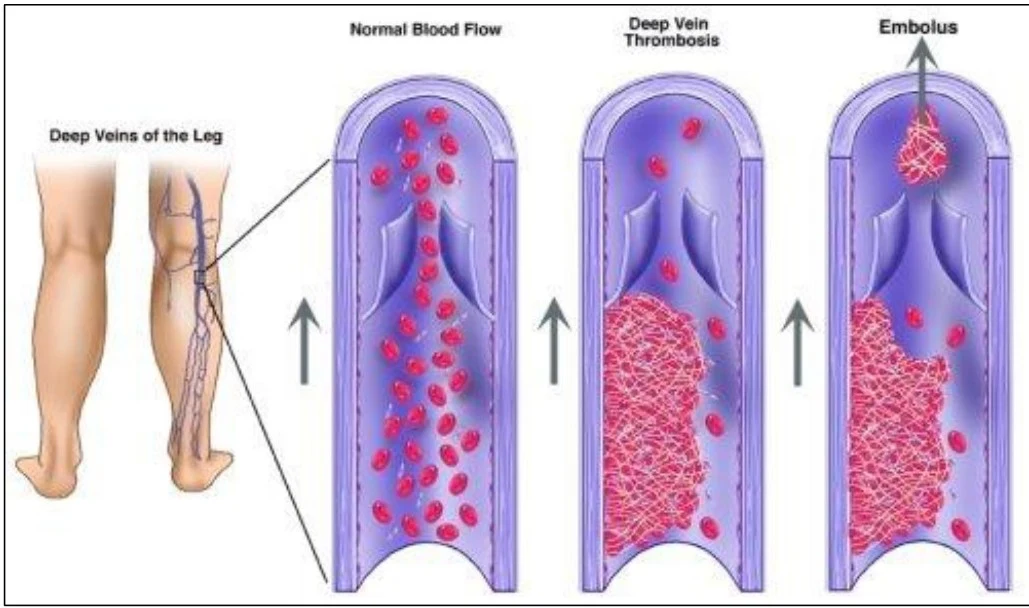
A thrombus is a clot that develops and stays within an unbroken blood vessel. If this clot breaks free and floats through the bloodstream, it is called an embolus. If an embolus obstructs a vessel, it causes an embolism, which can be life-threatening.
Diagram illustrating normal blood flow in the deep veins of the leg, the development of deep vein thrombosis (blood clot), and the formation of an embolus.
Keep reading for free.
Create an account to finish this reading and make it part of your study routine.