Biological Organization, Biomolecules, and Cellular Structures
An exploration of the human body's levels of organization, the four classes of biomolecules, cellular organelles, membrane transport, and primary tissue types.
Understanding the human body requires a journey from the microscopic to the macroscopic. To master anatomy and physiology, one must grasp how tiny atoms build complex molecules, how those molecules form the machinery of a cell, and how those cells organize into the tissues and organs that sustain life.
Levels of Biological Organization
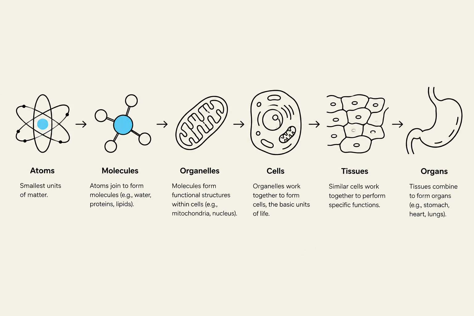
The human body is organized in a hierarchical fashion, moving from the simplest building blocks to the most complex systems. At the most fundamental level are atoms, which combine to form molecules. These molecules can serve as structural components or perform specific functions.
When molecules associate with one another, they form organelles—specialized subunits within a cell. Groups of similar cells working together constitute a tissue. When multiple tissues associate, they form an organ (such as the skin, stomach, or heart), and finally, these organs work together within organ systems to form the complete organism.
In medical study, we distinguish between gross anatomy, which focuses on large structures visible to the naked eye (like muscles and bones), and microanatomy, which requires a microscope to study structures like cells and tissues.
This diagram illustrates the hierarchical organization of life, starting from atoms and progressing through molecules, organelles, cells, tissues, and finally organs.
The Four Classes of Biomolecules
All biomolecules in the human body are carbon-based. They are constructed from smaller building blocks called monomers, which are assembled into larger polymers.
Carbohydrates: These are primary energy sources. Their monomers are ring-shaped monosaccharides (e.g., glucose). Larger chains, or polysaccharides, like glycogen, serve as energy storage in human cells.
Lipids: These include fats, oils, and steroids. Unlike other biomolecules, lipids do not have a single true monomer; they are typically composed of fatty acid chains and a glycerol backbone. They are hydrophobic, meaning they do not mix with water. Phospholipids are particularly vital as they form the basis of all animal cell membranes.
Proteins: The most diverse class of biomolecules, proteins are built from amino acids. They serve as structural components (like keratin), transporters in membranes, signaling hormones, and enzymes that catalyze chemical reactions.
Nucleic Acids: These include DNA, RNA, and ATP. Built from nucleotides, they store genetic information and facilitate energy transfer between cellular reactions.
Cellular Structure and Organelles
The cell is the basic unit of life. All cells contain cytoplasm, which includes the cytosol (the intracellular fluid containing water, nutrients, ions, and waste) and various specialized organelles.
The Control Center and Protein Synthesis
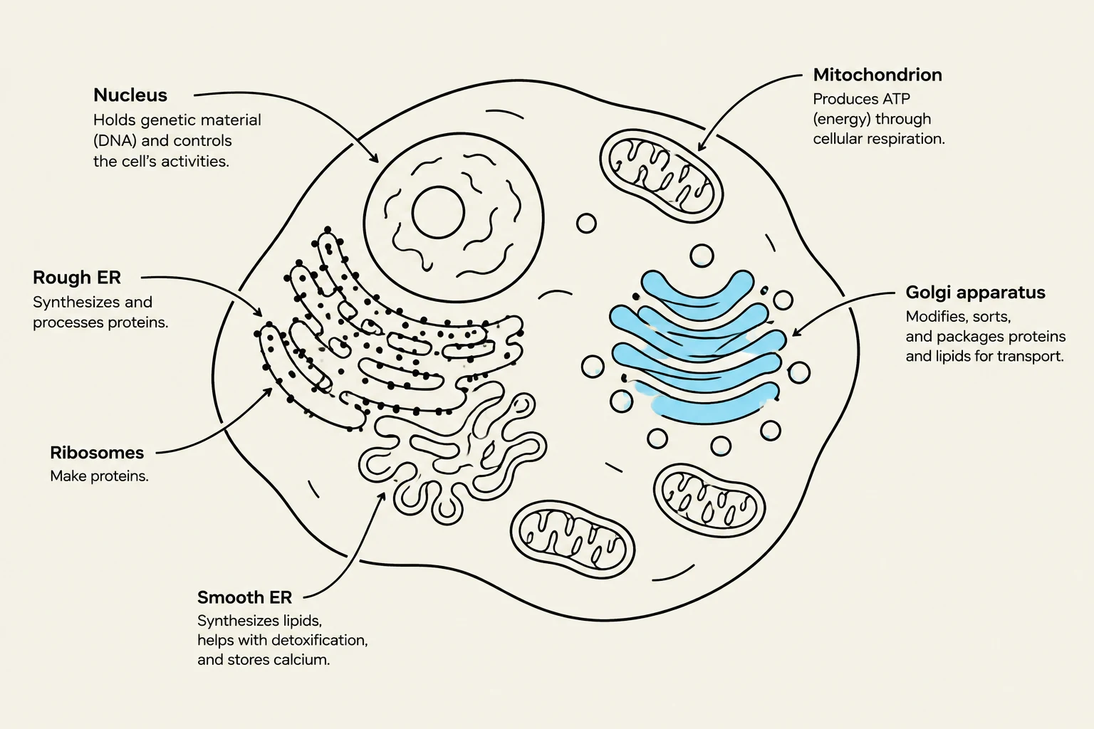
The nucleus acts as the cell's control center, housing the DNA. DNA provides the "blueprints" for protein synthesis. This process begins with transcription, where a gene from the DNA is copied into a messenger RNA (mRNA) molecule.
Next, ribosomes perform translation, using the mRNA instructions to assemble amino acids into a protein. Ribosomes exist in two forms:
Free ribosomes: Floating in the cytosol, producing proteins for use within the cell.
Fixed ribosomes: Attached to the rough endoplasmic reticulum (ER), producing proteins destined for secretion or for insertion into the cell membrane.
The smooth ER lacks ribosomes and specializes in synthesizing complex lipids and performing chemical detoxification. Once proteins are made by the rough ER, they are sent to the Golgi apparatus, a series of flattened sacs that modify, package, and distribute proteins via vesicles for export or internal use.
Energy and Support
The mitochondria are the "powerhouses" of the cell, performing aerobic respiration to produce ATP (adenosine triphosphate) using oxygen. If oxygen is unavailable, cells can produce ATP through anaerobic respiration (glycolysis) in the cytosol.
Structural integrity is provided by the cytoskeleton, a network of proteins that provides shape, resists pulling forces, and acts as internal "zip lines" for molecular transport. Additionally, cilia move fluids across the cell surface, while flagella allow the cell itself to move.
Diagram of an animal cell, illustrating key organelles such as the nucleus, mitochondria, endoplasmic reticulum, ribosomes, and Golgi apparatus, along with their functions.
Membrane Transport: Diffusion and Osmosis
The cell is enclosed by a selectively permeable membrane composed of a phospholipid bilayer. This membrane controls what enters and exits the cell.
Passive vs. Active Transport
Passive transport moves substances down their concentration gradient (from high to low concentration) without requiring energy.
Simple Diffusion: Small, hydrophobic molecules (like O2 or CO2) can pass directly through the lipid bilayer.
Facilitated Diffusion: Larger or charged molecules (like glucose or ions) require specific protein channels or carriers to cross.
Active transport moves substances against their concentration gradient (from low to high concentration) and requires energy in the form of ATP. A critical example is the sodium-potassium pump, which actively pumps three sodium (Na+) ions out of the cell and two potassium ($K^+$) ions into the cell.
Osmosis and Tonicity
Osmosis is the movement of water across a selectively permeable membrane to achieve equilibrium. Water movement is driven by the concentration of solutes; you can think of it as "water following salt".
To describe the effect of a solution on a cell, we use three terms:
Isotonic: The solute concentration is the same inside and outside the cell; there is no net movement of water.
Hypertonic: The solution has a higher solute concentration than the cell. Water leaves the cell, causing it to shrivel.
Hypotonic: The solution has a lower solute concentration than the cell. Water enters the cell, causing it to swell and potentially burst (lyse).
Exam Tip: Remember that the sodium-potassium pump moves 3 Na+ OUT and 2 K+ IN. A helpful mnemonic: "Na" has 3 letters (3 out) and "K" has 1 letter, but "potassium" has two "s" sounds or you can associate "K" with "In" because "K" is the 2nd letter of the word "In" (this is a common student trick, but focus on the 3:2 ratio!).
Vesicular Transport
For very large molecules, the cell uses vesicular transport:
Endocytosis: Bringing materials into the cell. This includes phagocytosis ("cell eating" of large particles) and pinocytosis ("cell drinking" of liquids).
Exocytosis: Expelling waste or products from the cell via vesicle fusion.
Histology: The Four Primary Tissue Types
Histology is the microscopic study of tissues. While a tissue is defined by its cell type, it may contain various specialized cells within that category.
1. Epithelial Tissue
Epithelial tissues line body surfaces and cavities and form glandular tissue. They possess several key characteristics:
Highly cellular: Cells are tightly packed with cellular junctions.
Polarity: They have an apical (unattached top) surface and a basal (attached bottom) surface.
Avascular: They contain no blood vessels; nutrients must diffuse from underlying tissues.
Regenerative: They have high rates of cell division to replace lost cells.
Epithelia are classified by the number of layers and cell shape. Simple epithelium (one layer) is optimized for rapid diffusion, such as simple squamous epithelium in the lungs. Stratified epithelium (multiple layers) provides protection, such as stratified squamous epithelium in the skin, which often contains keratin for strength and water resistance.
2. Muscle Tissue
Muscle tissue is specialized for contraction and movement:
Skeletal: Large, voluntary muscles responsible for gross body movements.
Cardiac: Found only in the heart; it is involuntary and self-pacing.
Smooth: Found in hollow organs (like the stomach or blood vessels); it is involuntary.
3. Connective Tissue
Connective tissues are characterized by having relatively few cells scattered within an extracellular matrix (ECM). All connective tissues derive from an embryonic tissue called mesenchyme. The ECM consists of protein fibers (made by cells like fibroblasts) and ground substance (inorganic components like calcium or water).
Dense Regular: Parallel fibers providing strength in one direction, found in tendons and ligaments.
Areolar: A loose, web-like tissue that is widely distributed for connection and support.
Supportive: Includes cartilage (for shock absorption) and bone (rigid, calcified support).
Specialized Cells: Includes fibroblasts (fibers), adipocytes (fat), osteocytes (bone), and chondrocytes (cartilage).
4. Neural Tissue
Neural tissue is responsible for rapid communication throughout the body. It consists of two primary cell types:
Neurons: The cells that actually send electrical signals.
Neuroglia: The "support crew" that cleans up extracellular fluid, maintains ion balance, and insulates axons to speed up signal transmission.
Diagrams illustrating the four main types of human tissue: epithelial, muscle, connective, and nervous tissue, with labels for their cellular components and functions.
Keep reading for free.
Create an account to finish this reading and make it part of your study routine.