Explore the structure and function of biological membranes, including the lipid bilayer, membrane proteins, and various transport mechanisms like diffusion and active transport.
Biological membranes are not merely passive boundaries; they are dynamic, selective, and highly organized structures that dictate the internal environment of a cell. By controlling the movement of ions, nutrients, and waste products, the membrane allows cells to maintain homeostasis, respond to signals, and perform specialized functions. Understanding how the molecular composition of the membrane influences its permeability and how various transport proteins move solutes is fundamental to physiology and medicine.
The Architecture of the Membrane
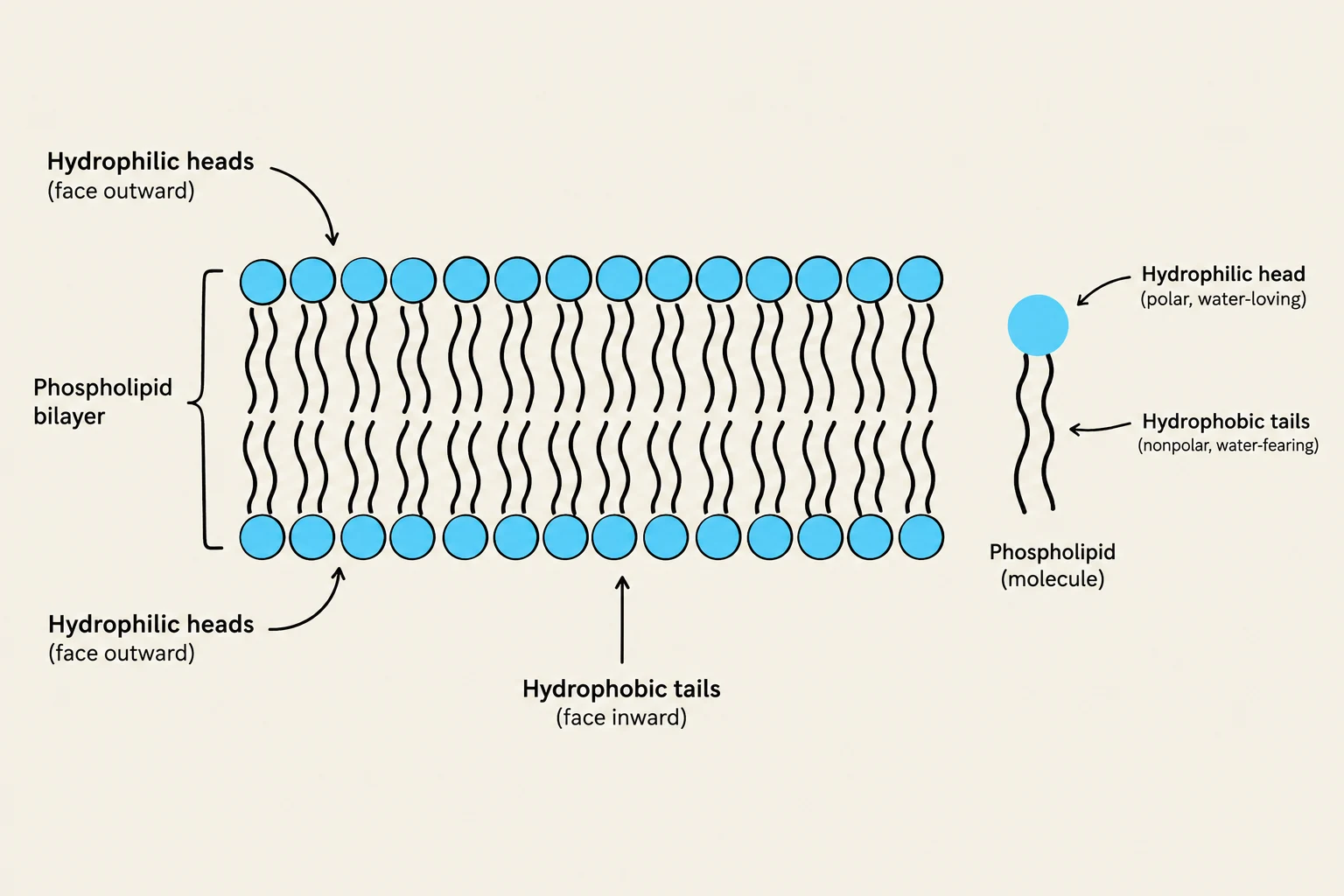
The foundation of the cell membrane is the lipid bilayer, composed primarily of amphipathic phospholipids. These molecules possess hydrophilic (water-loving) polar heads that face the aqueous environments of the extracellular fluid and cytoplasm, and hydrophobic (water-fearing) nonpolar tails that cluster together to form the membrane's interior core. This hydrophobic core acts as a formidable barrier to most water-soluble substances, such as ions and large polar molecules.
The physical properties of this bilayer are heavily influenced by its composition. For instance, the presence of unsaturated fatty acid tails prevents tight packing, thereby increasing membrane fluidity. Conversely, longer saturated tails increase packing and decrease fluidity. Cholesterol plays a critical role by acting as a buffer, stabilizing fluidity and reducing the permeability of the membrane to small, water-soluble molecules.
Beyond lipids, the membrane is reinforced by membrane-cytoskeleton attachments. Proteins like ankyrin and spectrin link integral membrane proteins to the internal cytoskeleton, providing mechanical stability and maintaining cell shape.
Clinical Correlation: Hereditary Spherocytosis
A defect in the genes encoding these attachment proteins (such as the ANK1 gene for ankyrin) can lead to hereditary spherocytosis. In this condition, red blood cells lose their normal biconcave shape and become spherical. These less deformable cells are easily trapped and destroyed in the spleen, leading to hemolysis and anemia.
Diagram illustrating the structure of a phospholipid bilayer, showing hydrophilic heads facing outward and hydrophobic tails facing inward, with a single phospholipid molecule depicted separately.
Principles of Diffusion and Fick's Law
Movement across the membrane can be passive (downhill) or active (uphill). Passive movement is driven by the electrochemical gradient, which is the sum of the concentration gradient (chemical force) and the electrical gradient (charge-based force). For ions, the electrical charge of the cell interior can either reinforce or oppose the concentration gradient.
The rate at which a molecule moves via diffusion is governed by Fick's Law. The net diffusion rate is determined by the permeability of the membrane, the surface area available, and the concentration gradient. This can be expressed mathematically as:
Rate=P⋅A⋅(Co−Ci)
Where:
P is permeability
A is surface area
Co and Ci are the concentrations outside and inside the cell, respectively.
Permeability (P) itself is defined by the diffusion coefficient (D), the partition coefficient (K), and the membrane thickness (Δx):
P=ΔxD⋅K
Clinical Application: Lung Disease
Fick's law explains why diffusion capacity falls in certain respiratory conditions. In pulmonary fibrosis, the diffusion barrier (Δx) becomes thicker, decreasing the rate of gas exchange. In emphysema, the destruction of alveolar walls reduces the available surface area (A), also lowering the diffusion rate.
Mechanisms of Passive Transport
Passive transport does not require direct energy (ATP) expenditure because solutes move down their electrochemical gradients. There are two primary modes of passive transport:
1. Simple Diffusion
Small, uncharged, or lipid-soluble molecules (such as O2 and CO2) can pass directly through the lipid bilayer. However, ions and large polar molecules like glucose have extremely low permeability through the lipid core alone.
2. Channel-Mediated Diffusion
Channels provide selective, aqueous pathways through the membrane. They allow specific ions to move rapidly down their electrochemical gradients. Channels are "gated," meaning they can open or close in response to voltage changes, ligands, or mechanical forces.
3. Carrier-Facilitated Diffusion
Unlike channels, which are open pores, carriers must bind to a specific solute. This binding induces a conformational change that exposes the solute to the opposite side of the membrane.
Because there are a finite number of carrier proteins, this mechanism is saturable. As the concentration of the solute increases, the transport rate eventually reaches a plateau known as the transport maximum (Vmax).
The Role of Insulin and GLUT Transporters
Glucose enters many cells via facilitated diffusion using GLUT transporters.
GLUT1/3: Found in RBCs and the brain; they provide basal glucose uptake and do not require insulin.
GLUT2: Found in the liver and beta cells; it allows for bidirectional glucose movement.
GLUT4: Found in skeletal muscle and adipose tissue. This transporter is insulin-responsive. When insulin binds to its receptor, it triggers the recruitment of more GLUT4 to the cell surface, increasing the cell's glucose-transport capacity.
Active Transport: Moving Against the Gradient
When a cell needs to move solutes "uphill" against their electrochemical gradient, it must use energy.
Primary Active Transport
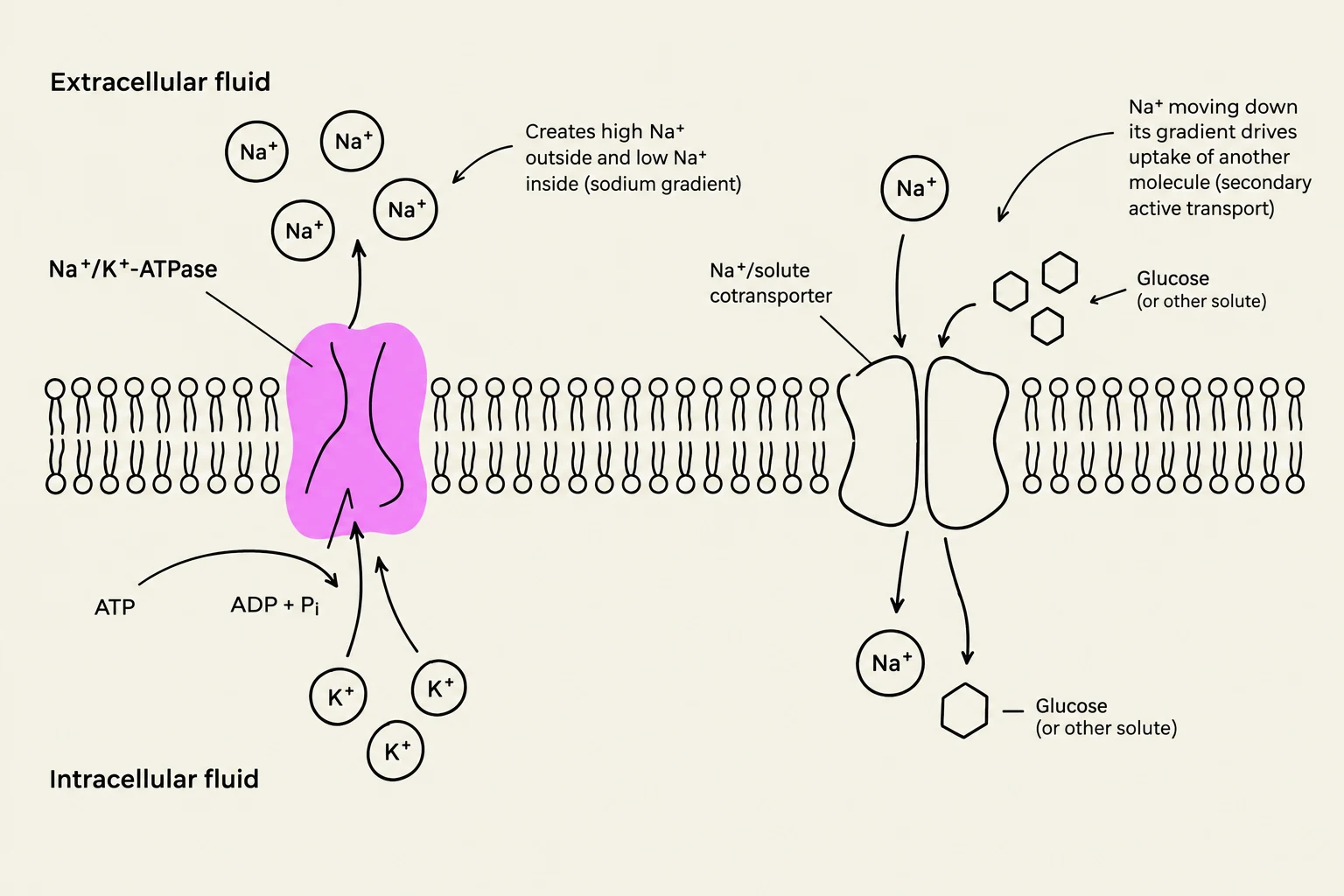
This mechanism is coupled directly to an energy source, most commonly the hydrolysis of ATP. A classic example is the Na+/K+-ATPase pump. For every ATP molecule hydrolyzed, the pump moves 3Na+ ions out of the cell and 2K+ ions into the cell. This process is electrogenic and is essential for maintaining the ion gradients that power other transport processes.
Secondary Active Transport
Secondary active transport does not use ATP directly. Instead, it uses the potential energy stored in an existing electrochemical gradient (usually the Na+ gradient created by the Na+/K+-ATPase) to drive another solute against its own gradient.
A prime example is the Na+-glucose cotransport (SGLT). In the small intestine (SGLT1) and the renal tubules (SGLT2), Na+ moves down its electrochemical gradient into the cell, "dragging" glucose along with it.
This diagram illustrates the sodium-potassium pump (Na+/K+-ATPase) and a sodium-glucose cotransporter embedded in a cell membrane. The Na+/K+-ATPase actively transports sodium ions out of the cell and potassium ions into the cell, maintaining a sodium gradient. This gradient is then used by the Na+/solute cotransporter to drive the uptake of glucose (or other solutes) into the cell, a process known as secondary active transport.
Clinical Case: Diabetic Ketoacidosis (DKA)
In a patient with Type 1 diabetes, the lack of insulin means GLUT4 cannot be recruited to the surface of muscle and fat cells, severely limiting glucose uptake. Meanwhile, the high blood glucose levels exceed the transport maximum of the kidney's SGLT transporters. This causes glucose to spill into the urine (glycosuria), which pulls water with it via osmosis, leading to dehydration and low blood pressure.
Summary Table of Transport Mechanisms
Pathway
Direction
Energy Source
Example
Simple Diffusion
Downhill
Concentration gradient
O2, CO2
Channel-Mediated
Downhill
Electrochemical gradient
$K^+$ through a channel
Carrier-Facilitated
Downhill
Concentration gradient
Glucose via GLUT transporters
Primary Active
Uphill
Direct ATP hydrolysis
Na+/K+-ATPase
Secondary Active
Uphill
Ion gradient (e.g., Na+)
Na+-glucose cotransport (SGLT)
End of Article
Keep reading for free.
Create an account to finish this reading and make it part of your study routine.