Basic Immunology and Host Defense: An Overview of the Immune Response
An overview of the human immune system, covering innate and adaptive immunity, pathogens, physical and biochemical barriers, inflammation, cellular recruitment, key immune cells, and the roles of cytokines.
The human body is constantly under siege by a diverse array of biological threats. To survive, we have evolved a sophisticated, multi-layered defense system designed to detect, neutralize, and remember these threats. This system, known as the immune system, is organized into distinct lines of defense that work in concert to maintain homeostasis and protect the host from disease.
The Nature of Pathogens
Before understanding how the body defends itself, we must define what it is defending against. A pathogen is any infectious agent that causes disease in its host. While many people associate pathogens exclusively with microbes, the term is broader.
Pathogens include:
Microbes: Such as viruses, bacteria, fungi, and prions.
Helminths: Parasitic worms that can cause significant disease.
It is important to note that not all microbes are harmful; for example, commensal bacteria (often called probiotics) live in our bodies without causing disease.
The First and Second Lines: Innate Immunity
The innate immune system is our evolutionarily older defense mechanism. It is characterized by being non-specific—meaning it responds to broad categories of threats rather than specific individual strains—and it provides an immediate response. However, because it does not tailor its response to a specific invader, it is generally not long-lasting.
The First Line: Physical and Biochemical Barriers
The primary goal of the first line of defense is to prevent infection from occurring in the first place. This is achieved through several types of barriers:
Physical/Mechanical Barriers: The skin and epithelium act as structural walls. Mechanical actions like sneezing, vomiting, or the sloughing of dead cells help physically expel invaders.
Biochemical Barriers: These include earwax, mucus, and various acids (such as sebaceous and lactic acids) that create hostile environments for microbes. Additionally, substances like surfactant help promote phagocytosis.
The Second Line: Inflammation and the Complement Cascade
If a pathogen breaches the physical barriers, the innate system moves into its second line of defense: inflammation. Inflammation is a generalized response designed to recruit immune cells to the site of infection. This process is characterized by:
Vasodilation (increased blood flow).
Increased capillary permeability (allowing fluid to move into tissues).
Swelling and the migration of cells like granulocytes and monocytes into the tissue.
During inflammation, the body also activates the complement cascade, a system of circulating plasma proteins that can directly destroy pathogens. One critical outcome of this cascade is the formation of the membrane attack complex, which creates pores in bacterial membranes, causing them to lyse.
Study Tip: Think of the innate immune system as the "first responders" at a crime scene. They arrive quickly and use standard procedures to contain the situation, but they don't necessarily know the specific identity of the perpetrator.
Sensing the Enemy: PRRs and PAMPs
How do immune cells "know" that a pathogen is present? They do not have eyes, but they do have specialized sensors. Immune cells express Pattern Recognition Receptors (PRRs). These receptors are designed to recognize Pathogen-Associated Molecular Patterns (PAMPs).
PAMPs are conserved molecular signatures—essentially "pieces" of infectious agents—that are found on fungal, bacterial, and viral agents but are not found in the host. The most famous family of PRRs are the Toll-like receptors (TLRs), of which there are at least ten known subtypes.
Cellular Recruitment: Chemotaxis and Extravasation
Once a pathogen is sensed, immune cells must travel from the bloodstream to the site of infection. This is achieved through leukocyte extravasation.
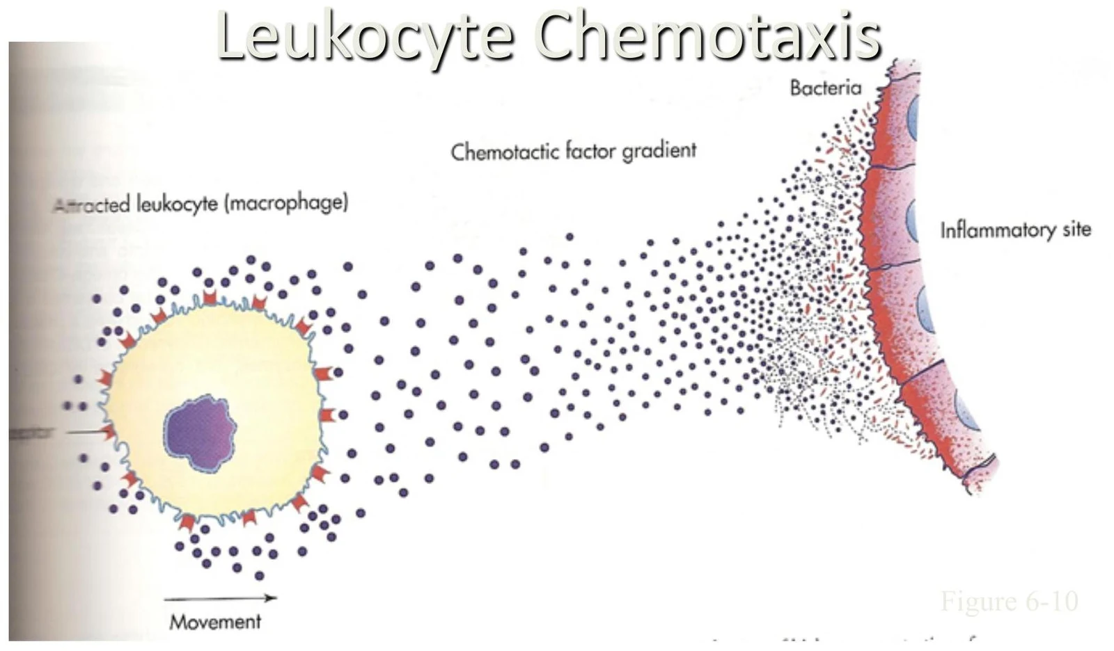
Chemotaxis: Leukocytes (white blood cells) use receptors to sense a high concentration of chemokines (chemical signals) at the infection site. They move toward this concentration gradient.
Diagram illustrating leukocyte chemotaxis, where a macrophage is attracted to an inflammatory site by a chemotactic factor gradient emanating from bacteria.
Extravasation Steps: The process involves several stages:
Margination and Pavementing: Cells move to the edge of the blood vessel and line up along the endothelial wall.
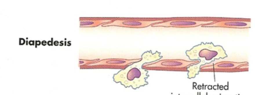
Diapedesis: The cells migrate through the endothelial lining to enter the tissue.
Diagram showing phagocytes in circulation within a blood vessel, illustrating endothelial cells and the basement membrane.Diagram illustrating the process of margination (pavementing) in a blood vessel, where white blood cells adhere to the vessel wall.Diapedesis is the movement of white blood cells from the bloodstream to the site of inflammation.Diagram illustrating exudation into inflamed tissue, showing cells moving through intercellular junctions.
Key Immune Cells
Various specialized cells perform different roles within the innate and adaptive systems:
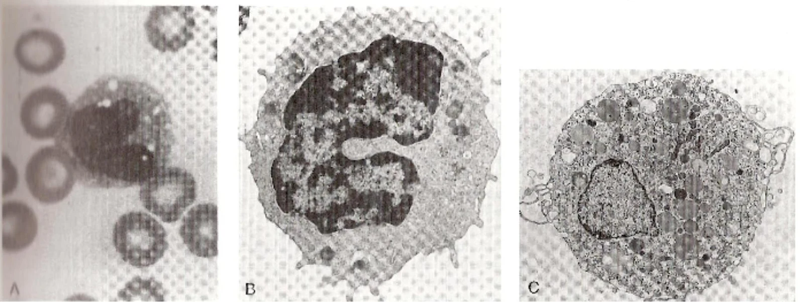
Macrophages/Monocytes: Monocytes circulate in the blood and migrate into tissues to become macrophages. They are professional phagocytes that engulf and ingest foreign material, using phagolysosomes to kill microbes with acids or reactive oxygen species (ROS).
Microscopic views of blood cells: A shows red blood cells and a monocyte. B and C are electron micrographs of white blood cells, likely neutrophils, illustrating their cellular structures.
Neutrophils (PMN): These are multi-lobular nucleus cells that arrive at the site of infection before macrophages. They are effective phagocytes but are relatively short-lived.
A micrograph shows a white blood cell, likely a neutrophil, surrounded by red blood cells.
Eosinophils: These cells are primarily involved in parasite defense and regulate mast cell mediators.
A micrograph of a neutrophil, a type of white blood cell, surrounded by red blood cells.
Mast Cells: These act as "sentinels" in the gut, lungs, and skin. When activated by stimuli like heat or microbes, they undergo degranulation, releasing histamine and other mediators.
The Third Line: Adaptive Immunity
The adaptive immune system is the third line of defense. Unlike the innate system, the adaptive response is highly specific to a particular pathogen and possesses immunological memory, allowing for a much faster and more effective response upon re-exposure.
T Cells and B Cells
Adaptive immunity is mediated by specialized lymphocytes:
T cells: Responsible for cell-mediated immunity. They use specific receptors to recognize pathogen markers.
B cells: Responsible for humoral (antibody) immunity. They produce antibodies that are specific to the invading pathogen.
Clonal Selection
The specificity of the adaptive response is driven by a process called clonal selection. The body produces a vast diversity of T and B cells, each with a unique receptor. When a specific antigen (a pathogen marker) enters the body, the individual T or B cell that possesses the matching receptor is "selected." This cell then undergoes rapid proliferation and differentiation to create a large army of identical cells specifically designed to fight that one pathogen.
Summary Table: Innate vs. Adaptive Immunity
Feature
Innate Immunity
Adaptive Immunity
Response Speed
Rapid/Immediate
Slower
Specificity
Non-specific/Generalized
Highly specific
Memory
No
Yes (Long-lived)
Lines of Defense
1st and 2nd
3rd
Orchestrating the Response: Cytokines
The entire immune response is coordinated by cytokines. These are chemical mediators produced by immune cells (or cells invaded by pathogens) that act as the "command and control" signals of the immune system. Much like catecholamines orchestrate the "fight-or-flight" response in the nervous system, cytokines orchestrate the inflammatory and adaptive responses.
Keep reading for free.
Create an account to finish this reading and make it part of your study routine.