This document describes immunochemistry methods for the detection and localization of proteins using antibodies, focusing on factors like antibody titer, dilution, incubation time, temperature, and enzyme substrate reactions.
Immunochemistry is a powerful scientific method used to detect and localize specific proteins or cellular components within a tissue sample using the highly specific binding properties of antibodies. In a clinical or research setting, this is most commonly applied through Immunohistochemistry (IHC). The ultimate goal of an IHC protocol is to achieve optimal specific staining—where the target protein is clearly visible—while simultaneously minimizing "background" interference, which can obscure results and lead to false interpretations.
The Fundamentals of Antibody Selection
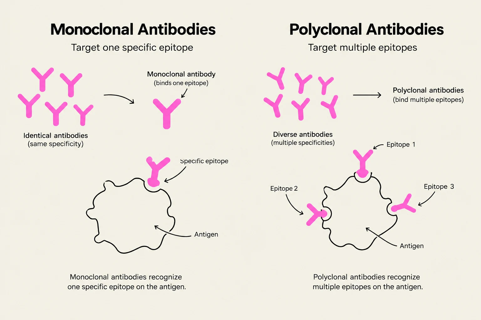
The success of an IHC procedure begins with the selection of the primary antibody. Antibodies generally fall into two categories: monoclonal antibodies and polyclonal antisera.
Monoclonal antibodies are derived from a single cell line and have a measurable, absolute concentration. Because they target a single epitope, they offer high specificity but can be sensitive to changes in the chemical environment.
Polyclonal antisera are a mixture of antibodies that recognize multiple epitopes on the same antigen. Unlike monoclonal antibodies, their strength is typically expressed as a "titer" determined through a series of serial dilutions.
Exam Tip: When comparing antibodies, remember that high-affinity antibodies (those that bind very strongly to the antigen) will generally react more quickly and produce more intense staining than low-affinity antibodies.
This diagram illustrates the difference between monoclonal and polyclonal antibodies. Monoclonal antibodies target a single specific epitope on an antigen, while polyclonal antibodies target multiple epitopes.
Optimizing the Staining Parameters
Achieving a perfect stain requires a delicate balance of several interdependent variables. If one parameter is changed, others must often be adjusted to compensate.
Antibody Titer and Dilution
The antibody titer is defined as the highest dilution that still yields maximum specific staining with minimal background. Finding this "sweet spot" is critical. Once the titer is known, the technician must apply the correct antibody dilution.
It is important to note that monoclonal antibodies are particularly sensitive to the pH and ionic strength of the diluent. For instance, a monoclonal antibody might show more intense staining if the pH is adjusted to 6.0 or if the concentration of Sodium Chloride (NaCl) is reduced.
Incubation Time and Temperature
The kinetics of the antigen-antibody reaction are heavily influenced by time and heat.
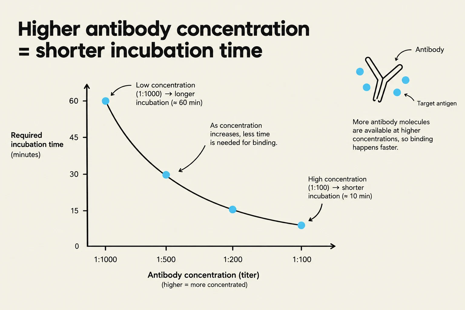
Time vs. Titer: There is an inverse relationship between these two; if you use a higher antibody titer (a more concentrated solution), you generally require a shorter incubation time.
Temperature: Increasing the temperature (for example, moving from room temperature to 37 °C) accelerates the reaction. However, higher temperatures require much stricter consistency in the laboratory environment, as even slight fluctuations can cause significant variations in staining intensity.
This graph illustrates the inverse relationship between antibody concentration and the required incubation time for binding. Higher antibody concentrations lead to shorter incubation periods, as more antibody molecules are available to bind with the target antigen.
Antigen Retrieval
Sometimes, the process of fixing tissue (such as using formalin) can "mask" the target protein, making it inaccessible to the antibody. To solve this, scientists use antigen retrieval (AR) techniques to unmask the epitopes. This can be achieved through:
Heat-Induced Epitope Retrieval (HIER): Using heat via autoclaves, pressure cookers, or microwaves.
Proteolytic Digestion: Using enzymes like pepsin to digest the protein matrix surrounding the antigen.
Enzymology and Signal Detection
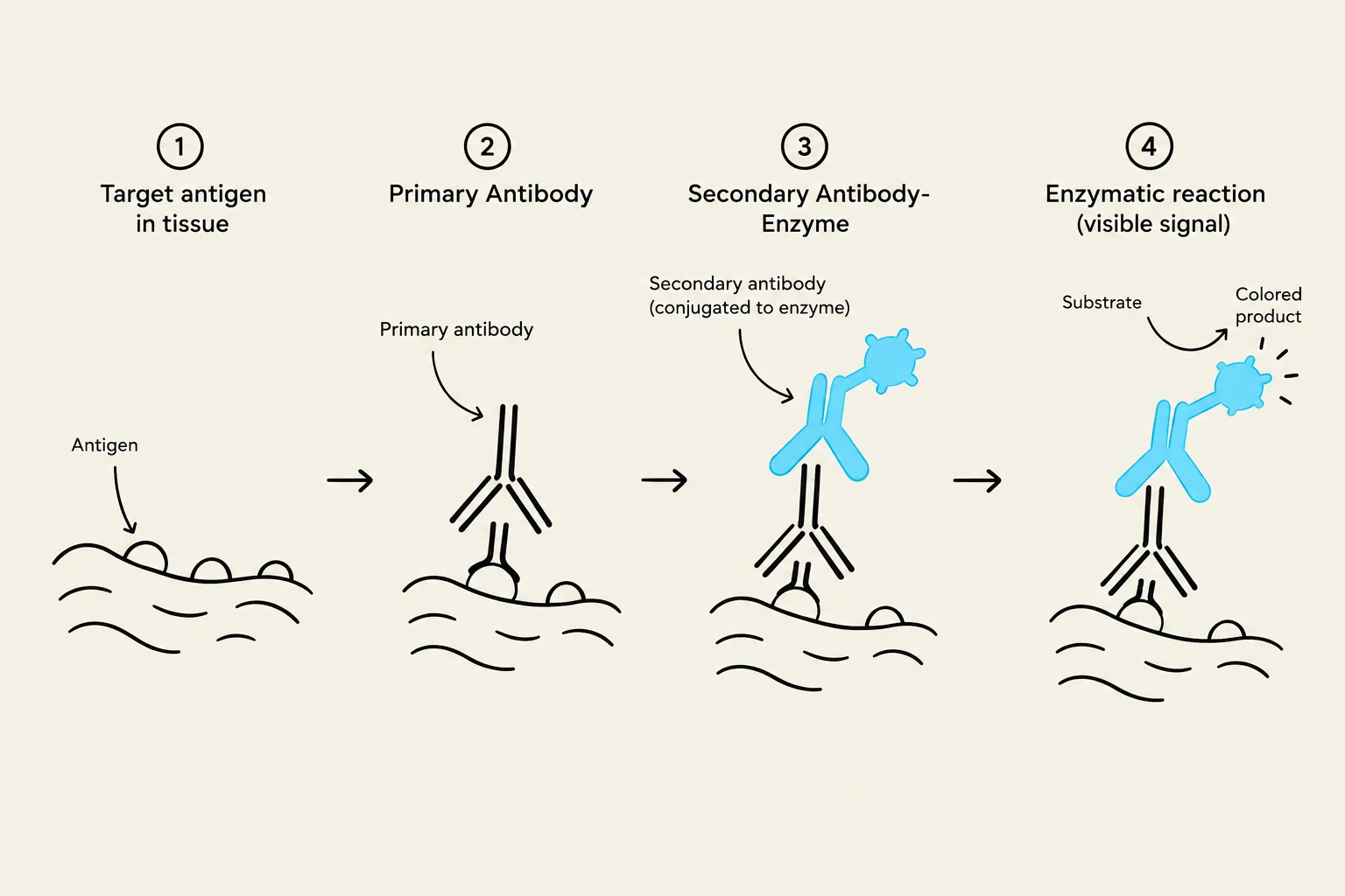
Once the antibody has bound to the target protein, we need a way to "see" it. This is achieved through immunoenzymatic methods, where an enzyme attached to the antibody reacts with a substrate-chromogen mixture to produce a visible color.
Horseradish Peroxidase (HRP)
HRP is a small enzyme (40 kDa) that catalyzes the oxidation of substrates using hydrogen peroxide (H2O2). The choice of chromogen (the molecule that provides the color) determines the appearance of the stain:
DAB (3,3'-diaminobenzidine): Produces a brown, highly insoluble product that is very stable.
AEC (3-amino-9-ethylcarbazole): Produces a rose-red color, but because it is alcohol-soluble, it requires the use of aqueous mounting media.
Alkaline Phosphatase (AP)
AP is a larger enzyme (100 kDa) that removes phosphate groups via hydrolysis. It is an excellent alternative when dealing with samples that have high endogenous enzyme activity (natural enzymes in the tissue that might interfere with the stain). For example, AP is preferred for blood and bone marrow smears because it avoids interference from endogenous peroxidase.
Common AP reactions include:
Naphthol AS-MX Phosphate: Used with chromogens like Fast Red TR (red) or Fast Blue BB (blue) to create soluble end products.
New Fuchsin: Produces an insoluble red end product, which is advantageous because it allows the specimen to be dehydrated in alcohol without losing the stain.
This diagram illustrates the steps of an immunoassay, showing how a primary antibody binds to an antigen, followed by a secondary antibody-enzyme conjugate, and finally a substrate reaction that produces a visible signal.
Summary Table: Enzyme Comparison
Feature
Horseradish Peroxidase (HRP)
Alkaline Phosphatase (AP)
Molecular Weight
≈40 kDa
≈100 kDa
Common Chromogen
DAB (Brown)
Fast Red (Red)
Key Advantage
Highly stable, insoluble
Avoids peroxidase interference
Key Limitation
Sensitive to endogenous peroxidase
Some products are alcohol-soluble
Keep reading for free.
Create an account to finish this reading and make it part of your study routine.