An introduction to mycology, covering fungal cell structure, nutrition, morphological classification (yeasts, molds, dimorphic fungi), reproduction, taxonomy, and clinical significance in veterinary medicine. It also discusses antifungal mechanisms and safety precautions.
Mycology is the study of fungi, a diverse group of eukaryotic, chemoorganotrophic, and non-motile organisms. Unlike bacteria, fungi possess membrane-bound organelles and a complex cell structure, including a rigid cell wall composed of chitinous fibrils embedded in a matrix of polysaccharides, proteins, and lipids. For veterinarians, understanding fungal biology is not merely academic; it is essential for diagnosing everything from common skin irritations to life-threatening systemic infections.
Fungal Cell Structure and Nutrition
Fungi obtain nutrients through extracellular digestion followed by absorption. Their growth is influenced by environmental factors: most fungi prefer an acidic pH (between 3.8 and 5.6) and an optimum temperature range of 25∘C to 30∘C for saprophytic (environmental) species. Parasitic fungi, however, often thrive at higher temperatures, typically 30∘C to 37∘C.
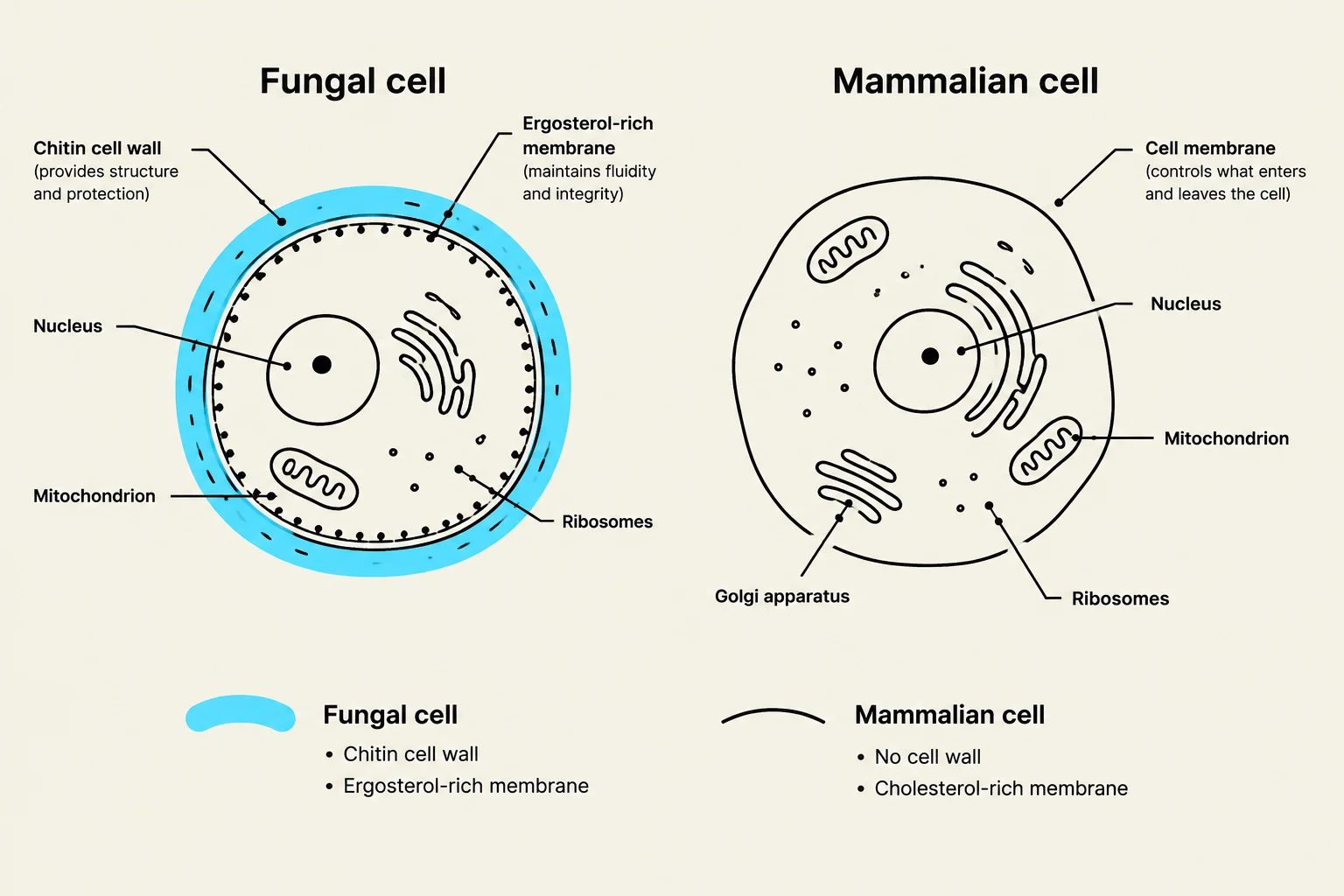
A defining feature of the fungal plasma membrane is the presence of ergosterol. While mammalian membranes rely on cholesterol, fungi use ergosterol to maintain membrane integrity. This biochemical difference is the "Achilles' heel" of fungi and serves as the primary target for many antifungal medications.
Diagram comparing the structures of a fungal cell and a mammalian cell, highlighting key organelles and cell wall differences.
Morphological Classification
Fungi are classified by their vegetative structure, known as a thallus. They generally fall into three morphological categories:
1. Yeasts
Yeasts are unicellular, round, or oval bodies. They primarily reproduce via an asexual process called budding, where a protuberance develops and eventually separates from the parent cell. The spores produced during this process are known as blastoconidia. Some fungi, like Candida albicans, exhibit "yeast-like" growth, forming chains of elongated cells called pseudohyphae when the buds fail to detach.
2. Filamentous Fungi (Molds)
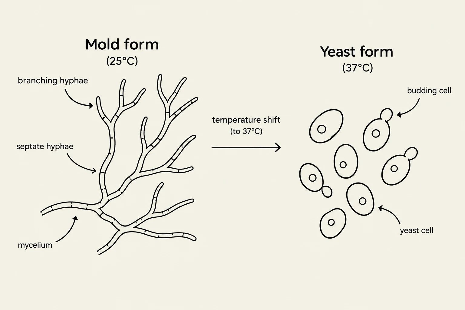
Molds are multicellular and composed of branching filaments called hyphae. A mass of intertwined hyphae is referred to as a mycelium. Hyphae are categorized by their structure:
Specialized hyphae include vegetative (nutrient-absorbing), aerial (growing above the surface), and reproductive (bearing spores). Certain dermatophytes produce unique hyphal shapes, such as spiral, pectinate (comb-like), or racquet-shaped hyphae, which are vital for laboratory identification.
3. Dimorphic Fungi
Dimorphic fungi are significant veterinary pathogens that can switch between forms based on temperature—a phenomenon known as the Yeast-to-Mold (YM) shift. They typically exist as molds in the environment (25∘C to 30∘C) and convert to yeast in host tissues (30∘C to 37∘C). Examples include Histoplasma, Blastomyces, and Coccidioides.
This diagram illustrates the dimorphism of certain fungi, showing their transition from a mold form at 25°C, characterized by branching and septate hyphae forming a mycelium, to a yeast form at 37°C, where individual yeast cells reproduce via budding.
Reproduction and Spore Types
Fungi reproduce through both asexual and sexual methods.
Asexual reproduction is the most common method for pathogens and involves several spore types:
Sporangiospores: Produced within a sac-like structure called a sporangium.
Conidia: Small spores attached to a specialized stalk called a conidiophore.
Arthrospores: Formed by the fragmentation of hyphae.
Sexual reproduction involves the fusion of gametangia (plasmogamy) and the subsequent fusion of nuclei (karyogamy). This results in specialized spores such as ascospores (in sac fungi like Aspergillus), basidiospores (in club fungi like mushrooms), and zygospores.
Taxonomy and Clinical Mycoses
Fungi are taxonomically grouped into several classes:
Ascomycota (Sac Fungi): Septate fungi that produce ascospores (e.g., Aspergillus, Penicillium).
Basidiomycota (Club Fungi): Highly developed, septate mycelia that produce basidiospores (e.g., mushrooms).
Deuteromycetes (Fungi Imperfecti): A group containing many pathogens, such as dermatophytes, that lack a known sexual cycle.
Classification of Mycoses
Clinical fungal infections, or mycoses, are categorized by the depth of tissue invasion:
Superficial/Cutaneous: Confined to the outermost layers of skin, hair, and nails. Dermatophytes are the primary agents here, as they have the ability to destroy keratin.
Subcutaneous: Involve deeper tissues, such as muscle.
Systemic: The most serious form, where fungi invade deep tissues and organs, often causing life-threatening symptoms.
Exam Tip: When discussing systemic mycoses like Histoplasma or Blastomyces, always consider the patient's geography and travel history. These diseases are often endemic to specific regions (e.g., Histoplasma in the Ohio/Mississippi River valleys).
Veterinary Clinical Significance and Safety
Fungi can be saprophytes (living on organic matter), opportunists (infecting compromised hosts), or zoonotic (transmissible from animals to humans). For example, Microsporum canis is a common dermatophyte in cats that can readily infect humans.
Safety Warning: When handling suspected fungal samples, particularly those that may be dimorphic, always wear gloves. Some molds produce highly infectious aerosols; avoid opening suspect cultures outside of appropriate containment to prevent accidental inhalation.
This diagram illustrates the process of diagnosing a fungal infection, starting with clinical history and geography, followed by sample collection (skin scraping or hair), laboratory analysis (culture, microscopy, or molecular testing), and finally diagnosis.
Antifungal Mechanisms
Because fungi are eukaryotic, finding drugs that kill them without harming the host is challenging. Most antifungals target the cell membrane or wall:
Polyenes (e.g., Amphotericin B): Bind directly to ergosterol to create pores in the membrane, causing leakage.
Azoles (e.g., Itraconazole, Fluconazole): Inhibit the synthesis of ergosterol.
Allylamines (e.g., Terbinafine): Block an early step in ergosterol production, leading to toxic buildup of squalene.
Echinocandins: Target the β-1,3-glucan in the cell wall.
Keep reading for free.
Create an account to finish this reading and make it part of your study routine.