


Understanding how the heart functions requires a dual perspective: the mechanical action of the muscle and the electrical signals that trigger that action. An electrocardiogram (ECG) is a specialized recording of the heart's electrical activity. By monitoring the movement of ions across cell membranes, an ECG allows clinicians to assess vital information, including heart rate, rhythm, axis, hypertrophy, and signs of infarction.
The Cellular Basis of Cardiac Activity
The heart is composed of various specialized cells, but two types are critical for understanding electrical conductivity and contraction: myocardial cells and conduction cells.
Myocardial cells (or contractile cells) are responsible for the kinetic work of the heart. These cells are located in the atrial and ventricular walls and must receive electrical stimulation to contract. Within these cells, myofibrils containing actin and myosin filaments slide together to facilitate contraction.
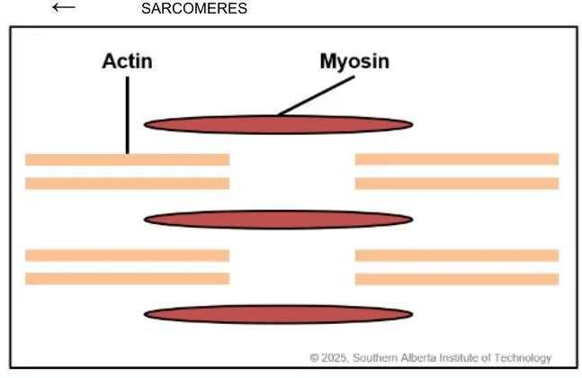
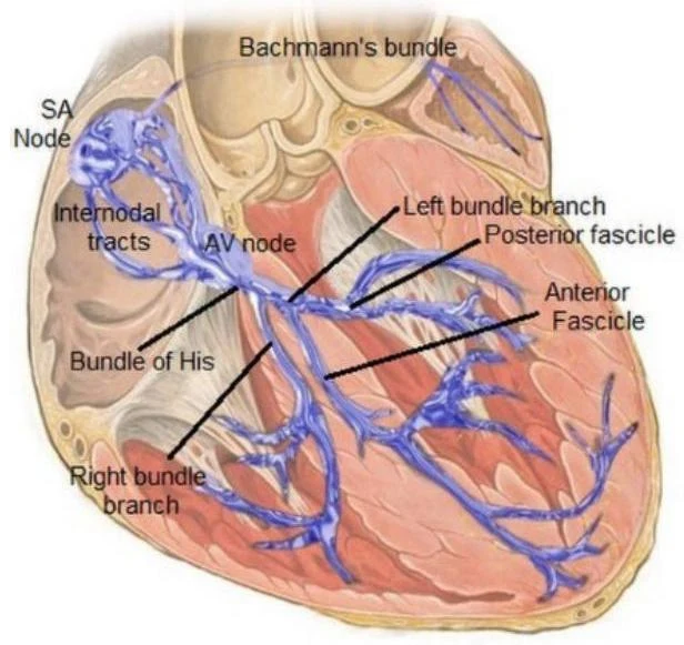
Conduction cells (or auto-arrhythmic cells) are specialized to initiate and transmit electrical impulses throughout the heart, though they do not possess the ability to contract.
Depolarization and Repolarization
The electrical signal of the heart is an "action potential"—a wave of electrical discharge caused by the movement of ions (specifically Sodium, Potassium, and Calcium) across the cell membrane. This movement is driven by electrochemical gradients.
- Depolarization: This is the process where a cell becomes positively charged. In the heart, depolarization is the trigger for myocardial contraction.
- Repolarization: This is the recovery phase where the cell returns to its resting, negative state (polarized).
Keep reading for free.
Create an account to finish this reading and make it part of your study routine.
Continue reading freeAlready have an account? Log in
Updated