An overview of basic cardiac anatomy, including the heart's chambers, valves, and the pathway of blood flow through the circulatory system. It also covers arteries, veins, and coronary circulation.
Understanding the cardiovascular system is fundamental to clinical practice. The heart acts as a sophisticated dual-pump system, driving blood through a continuous loop of circulation to ensure every cell in the body receives oxygen and nutrients while removing metabolic waste. To master cardiac diagnostics, one must first master the underlying anatomy, the direction of blood flow, and the physiological mechanisms that regulate vascular diameter.
Cardiac Anatomy: Chambers and Valves
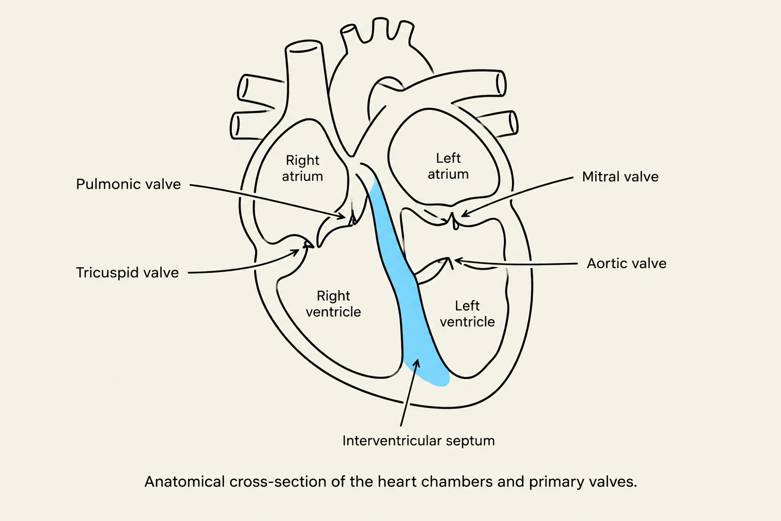
Anatomical cross-section of the heart chambers and primary valves.
The heart is a muscular organ divided into four distinct chambers that work in coordination to facilitate blood movement. These chambers are categorized into two upper receiving chambers (atria) and two lower pumping chambers (ventricles).
The Four Chambers
Right Atrium: Receives deoxygenated blood from the body.
Right Ventricle: Pumps deoxygenated blood toward the lungs.
Left Atrium: Receives oxygenated blood returning from the lungs.
Left Ventricle: The most muscular chamber, responsible for pumping oxygenated blood to the entire body.
A critical structural component is the septum, a muscular wall that separates the left and right sides of the heart. The septum is vital because it prevents the mixing of oxygenated and deoxygenated blood.
Heart Valves
To ensure that blood flows in only one direction (unidirectional flow), the heart utilizes four primary valves:
Tricuspid Valve: Located between the right atrium and right ventricle.
Pulmonic Valve: Located between the right ventricle and the pulmonary artery.
Mitral Valve: Located between the left atrium and left ventricle.
Aortic Valve: Located between the left ventricle and the aorta.
The Pathway of Blood Flow
Diagram illustrating the pulmonary and systemic circulation of blood through the heart, lungs, and body.
Blood follows a highly specific physiological sequence to ensure efficient oxygenation:
Deoxygenated Circuit: Deoxygenated blood enters the right atrium, moves into the right ventricle, and is then pumped through the pulmonary artery to the lungs.
Oxygenation: In the lungs, blood picks up oxygen.
Oxygenated Circuit: Oxygenated blood returns to the heart via the pulmonary vein into the left atrium, moves into the left ventricle, and is finally ejected through the aorta to supply the rest of the body.
Vascular Circulation: Arteries, Veins, and Coronary Flow
The circulatory system is composed of vessels categorized by the direction of blood flow relative to the heart.
Arteries vs. Veins
Arteries: These vessels carry blood away from the heart. In the systemic circuit, arteries typically carry oxygenated blood.
Veins: These vessels carry blood to the heart. In the systemic circuit, veins typically carry deoxygenated blood from the extremities back to the heart to be replenished with oxygen.
Coronary Circulation
While the heart pumps blood to the body, it also requires its own dedicated blood supply to function. This is known as coronary circulation. The coronary arteries supply oxygen directly to the heart muscle (myocardium). Key arteries include:
Left Main Coronary Artery
Left Anterior Descending (LAD)
Circumflex Artery
Right Coronary Artery (RCA)
Clinical Alert: Obstructions or blockages in these coronary vessels can lead to ischemia (restricted blood supply) or infarction (tissue death/myocardial infarction).
Regulation of Vessel Diameter
Comparison of vasoconstriction and vasodilation in blood vessels.
The body regulates blood pressure and flow by changing the diameter of the blood vessels through two primary processes:
Process
Action
Common Triggers
Vasoconstriction
Narrowing of blood vessels
Cold, stress, nicotine, vasopressors
Vasodilation
Widening of blood vessels
Heat, exercise, alcohol, nitrates
Diagnostic Review: Laboratory and Imaging
When cardiac dysfunction is suspected, clinicians rely on a combination of laboratory markers and diagnostic imaging to assess the heart's health.
Cardiac Laboratory Markers
Laboratory tests provide biochemical evidence of cardiac stress or damage:
Troponin: The most sensitive indicator of myocardial damage. It helps differentiate cardiac from non-cardiac chest pain. It typically rises 4–6 hours after an MI, peaks at 10–24 hours, and returns to baseline in 10–14 days.
CK-MB: An older marker of muscle damage; it rises 6 hours after an MI and peaks at 18 hours.
BNP / Pro-BNP: These are released from the cardiac ventricles. BNP is used to distinguish cardiac from respiratory causes of shortness of breath (dyspnea), often seen in Congestive Heart Failure (CHF). Pro-BNP is more specific to Left Ventricular dysfunction.
Inflammatory Markers (CRP & ESR): C-Reactive Protein (CRP) and Erythrocyte Sedimentation Rate (ESR) indicate inflammation or infection.
Noninvasive Imaging and Procedures
EKG/ECG (12-lead): Provides information regarding the electrical conduction of the heart.
Holter Monitor: A continuous 24-hour monitor used to diagnose arrhythmias like atrial fibrillation or causes of fainting.
Echocardiogram: Uses sound waves to create a moving picture of the heart to evaluate valves and chambers.
Ejection Fraction (EF): A key measurement from an echo that indicates the effectiveness of the heart's "pump." Normal values are 55–70%; values under 40% are often seen in Heart Failure.
Bubble Study: Used during an echo to detect a Patent Foramen Ovale (PFO), which is a tiny opening between the right and left atria.
Stress Test: Evaluates the heart's response to physical exertion (treadmill or bike) to unmask symptoms of Coronary Artery Disease (CAD).
Invasive and Specialized Diagnostics
Angiography: Uses radiopaque contrast medium and X-rays to visualize vessels.
CT/MR Angiography: CT uses multilayered 3D imaging, while MR uses magnetic resonance and does not rely on radiation or iodine-based dye.
Duplex/Doppler Ultrasound: Measures the speed and diameter of blood flow through vessels.
Clinical Calculation: Ankle-Brachial Index (ABI)
The Ankle-Brachial Index (ABI) is a non-invasive screening tool used to detect Peripheral Artery Disease (PAD) by comparing the blood pressure in the ankle to the blood pressure in the arm.
The Formula
The ABI is calculated as:
ABI=Systolic Pressure in ArmSystolic Pressure in Ankle
Interpretation of Results
Normal:0.9−1.3
Claudication (Leg pain during walking):0.4−0.9
Occlusive Arterial Disease/Severe PAD:<0.4 (often associated with non-healing ulcers or ischemic rest pain)
Practice Example:
A patient has a Right Brachial Systolic pressure of 125 mm and a Right Ankle Systolic pressure of 75 mm.
ABI=12575ABI=0.6
Result: This patient's ABI is 0.6, which falls into the range often associated with claudication.
Keep reading for free.
Create an account to finish this reading and make it part of your study routine.