


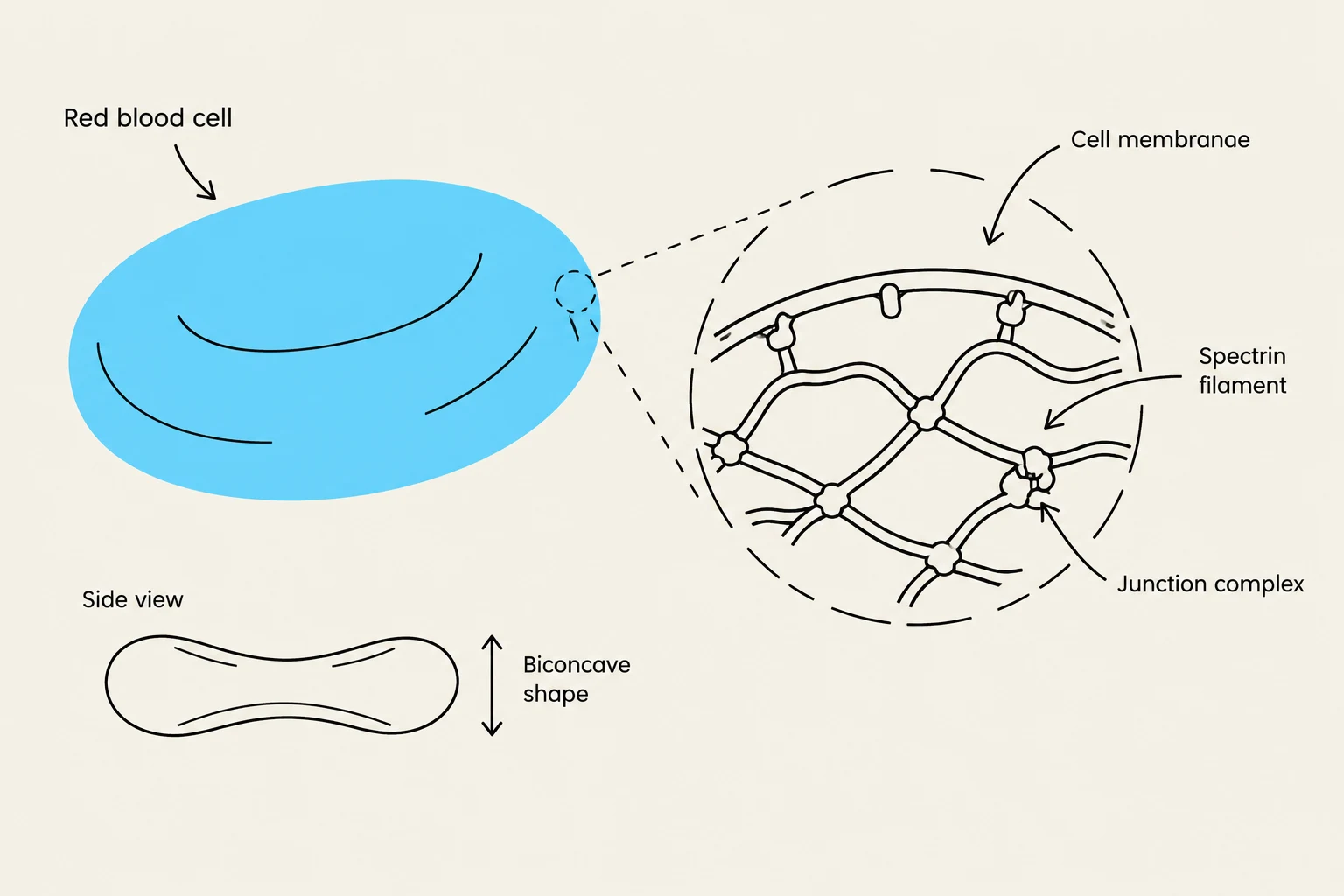
Cell membranes are far more than simple boundaries; they are dynamic, highly organized structures that facilitate energy production and maintain physical shape. To understand how a cell functions, one must look at both the molecular machines embedded within the membrane and the structural scaffolding that supports it. This article explores two distinct but fundamental biological systems: the light-driven proton pump known as bacteriorhodopsin and the protein meshwork of the cell cortex.
Bacteriorhodopsin: A Light-Driven Energy Converter
In certain microorganisms, the ability to convert light into chemical energy is facilitated by a specialized transmembrane protein called bacteriorhodopsin. This protein acts as a proton pump, a mechanism that moves ions across a membrane to create a concentration gradient.
The architecture of bacteriorhodopsin is highly specific. It is composed of approximately 250 amino acids arranged into seven helices. These helices are embedded vertically within the hydrophobic interior of the lipid bilayer. The "engine" of this protein is a small molecule called retinal, which is positioned across the transmembrane strands.
The Mechanism of Proton Pumping
The process of energy conversion follows a precise sequence of structural changes:
- Photon Absorption: When retinal absorbs a photon of light, it undergoes a rapid conformational change.
- Conformational Shift: This change in retinal triggers a series of structural shifts in the surrounding seven helices.
- Proton Translocation: These shifts create a pathway through the protein. Strategically placed polar amino acid side chains act as stepping stones, allowing a proton ($H^+$) to bypass the hydrophobic lipid environment and move from the cytosol to the extracellular space.
- Regeneration: Once the proton is released outside, the retinal is regenerated by taking up a new $H^+$ from the cytosol, resetting the pump for the next cycle.
Keep reading for free.
Create an account to finish this reading and make it part of your study routine.
Continue reading freeAlready have an account? Log in
Updated