This guide explains the essential biochemical and physiological tests used in microbiology laboratories to identify bacterial species, focusing on Gram-positive and Gram-negative bacteria.
In the microbiology laboratory, identifying a pathogen goes far beyond simply seeing a cell under a microscope. While Gram staining provides a foundational starting point by revealing cell wall structure and morphology, clinicians must use a series of biochemical and physiological tests to pinpoint the exact species responsible for an infection. This process involves observing how bacteria interact with specific nutrients, how they respond to antibiotics, and how they produce certain enzymes.
Morphological Foundations: Cocci and Arrangement
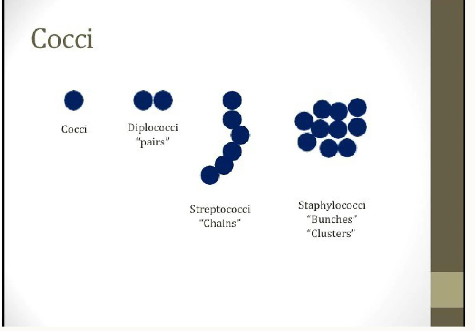
Before performing biochemical tests, microbiologists examine the physical arrangement of bacteria. For cocci (round bacteria), the way they stick together is a primary diagnostic clue.
Diagram illustrating different arrangements of cocci bacteria: single cocci, diplococci (pairs), streptococci (chains), and staphylococci (clusters).
Diplococci: Bacteria appearing in pairs.
Streptococci: Bacteria arranged in long chains.
Staphylococci: Bacteria appearing in grape-like "bunches" or clusters.
Microscopic view of Streptococci bacteria arranged in chains.
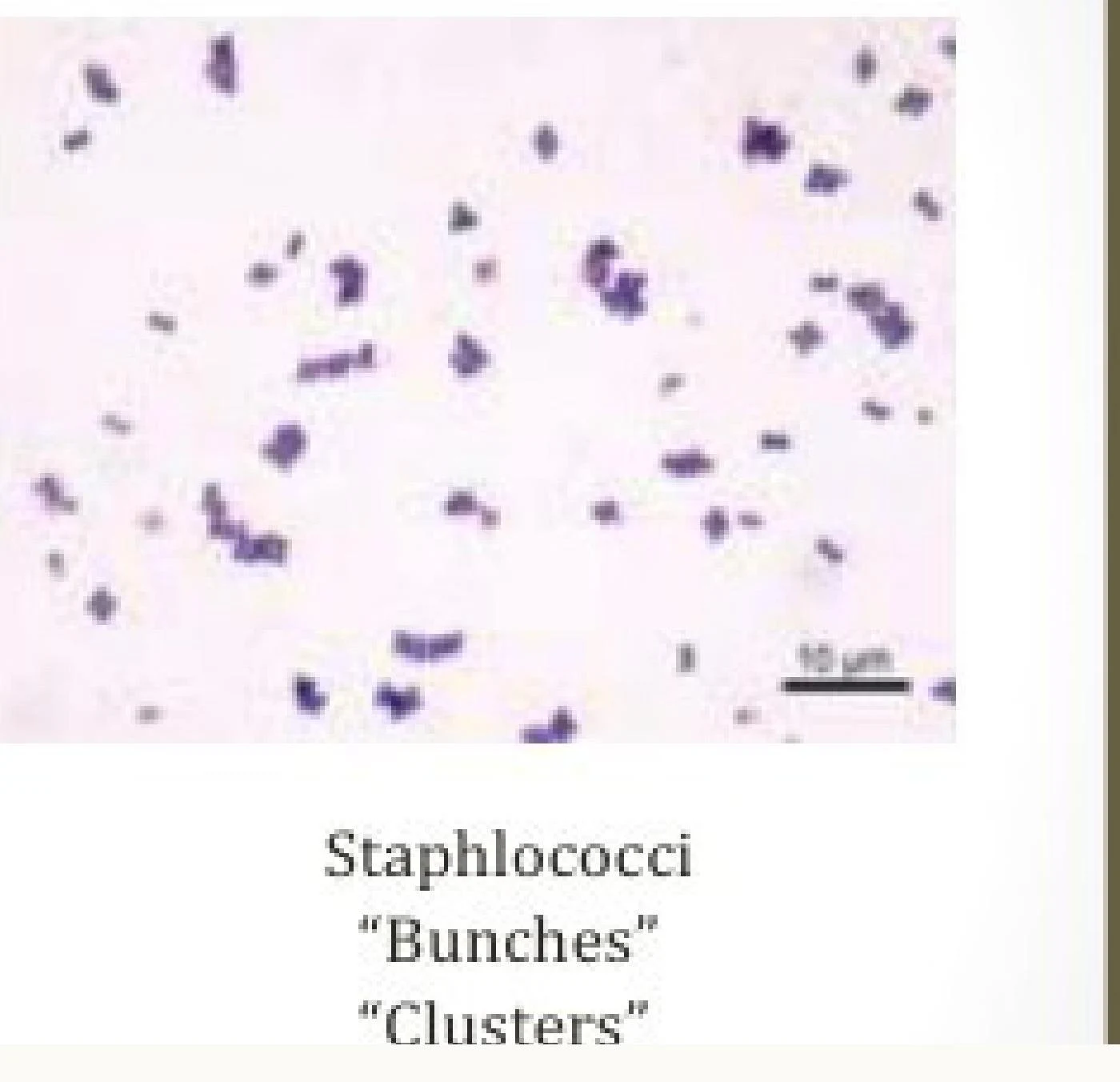
Microscopic view of Staphylococci bacteria, characterized by their arrangement in bunches or clusters.
Identifying Gram-Positive Cocci
Once the arrangement is noted, a series of tests helps differentiate between the major families of Gram-positive cocci.
The Catalase Test: Staph vs. Strep
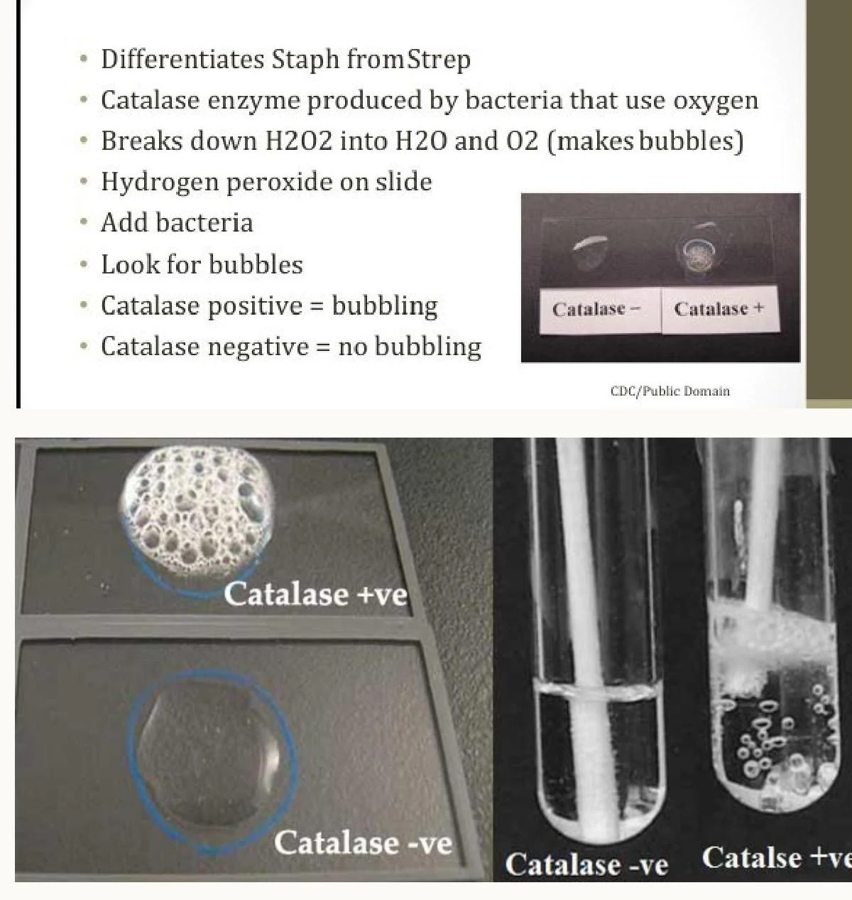
The first major branch in the diagnostic roadmap is the catalase test. Catalase is an enzyme produced by bacteria that use oxygen; it breaks down hydrogen peroxide (H2O2) into water (H2O) and oxygen gas (O2). When hydrogen peroxide is added to a bacterial sample, the presence of bubbles indicates a positive result.
This image illustrates the catalase test, a method used to differentiate bacteria like Staphylococcus (catalase-positive, showing bubbling) from Streptococcus (catalase-negative, no bubbling) by observing the reaction of hydrogen peroxide with bacterial enzymes.
Positive (Bubbles):Staphylococcus species.
Negative (No bubbles):Streptococcus species.
Differentiating Staphylococcus Species
If the catalase test is positive, the next step is to differentiate between various Staphylococcus species using the Coagulase Test and Mannitol Salt Agar (MSA).
The coagulase test determines if a bacterium produces an enzyme that causes rabbit plasma to clot.
Positive (Clumping):Staphylococcus aureus.
Negative (No clumping): Other species, such as S. epidermidis or S. saprophyticus.
To further refine this, Mannitol Salt Agar (MSA) is used. This medium is selective because its high salt concentration (7.5%) inhibits most other bacteria, but Staphylococci can tolerate it. It is also differential: if the bacteria ferment the sugar mannitol, they produce acid, which turns the phenol red indicator in the agar from red to yellow.
Yellow colonies:Staphylococcus aureus (a mannitol fermenter).
Pink/Red colonies: Other Staph species (non-fermenters).
For the specific differentiation of S. saprophyticus from S. epidermidis, clinicians use Novobiocin resistance testing. By measuring the zone of clearance around a Novobiocin antibiotic disk, they can identify the resistant S. saprophyticus.
Differentiating Streptococcus Species
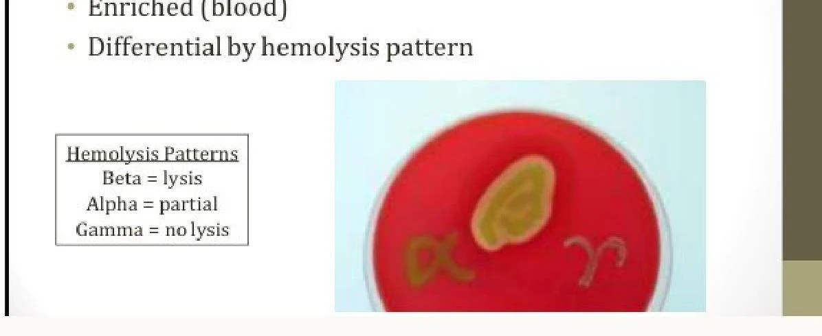
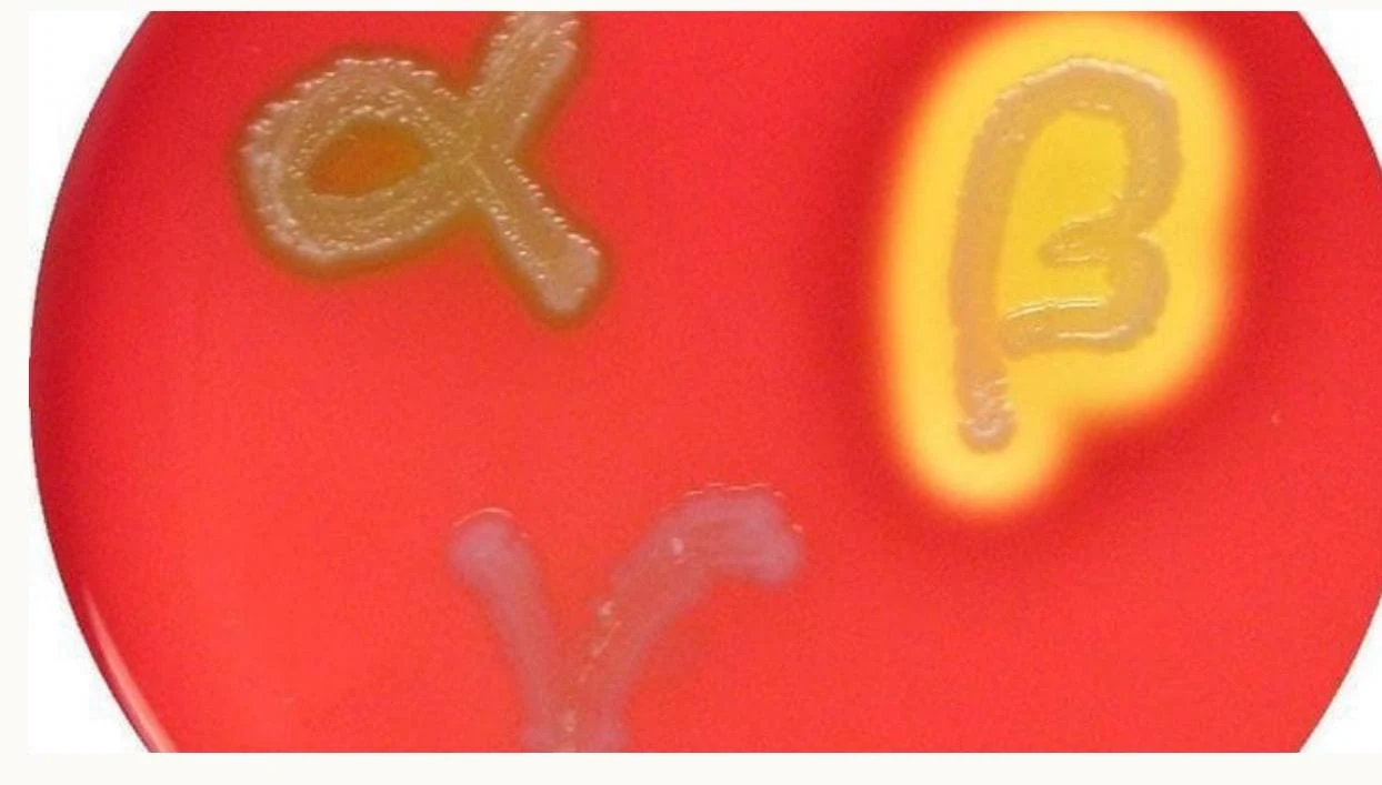
If the catalase test is negative, the focus shifts to the Streptococcus genus. This often begins with observing hemolysis patterns on Blood Agar.
This image shows different types of hemolysis patterns observed on a blood agar plate, including beta (lysis), alpha (partial lysis), and gamma (no lysis).
Bacterial cultures labeled with Greek letters alpha and beta on a red agar plate.
Beta (β): Complete lysis of red blood cells.
Alpha (α): Partial lysis.
Gamma (γ): No lysis.
Specific antibiotic susceptibility tests are then used to categorize these groups:
Bacitracin: Differentiates Group A (S. pyogenes) from Group B (S. agalactiae). Group A is sensitive (large clearance zone), while Group B is resistant.
Optochin: Differentiates S. pneumoniae from S. viridans. S. pneumoniae is highly sensitive to Optochin.
The Group D Workflow
For Group D Streptococci (such as Enterococcus and S. bovis), a specific two-step sequence is used:
Step 1: Bile Esculin Agar. This medium is selective for Group D. If the bacteria hydrolyze esculin, the media turns dark brown or black.
Step 2: NaCl Media. To distinguish Enterococcus from S. bovis, bacteria are grown in high-salt media. Enterococcus is salt-tolerant and will grow, whereas S. bovis will not.
Identifying Gram-Negative Bacteria
Gram-negative identification often relies on how bacteria process carbohydrates and their enzymatic capabilities.
MacConkey's Agar and Lactose Fermentation
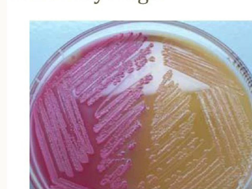
MacConkey's Agar is a primary tool for Gram-negative identification. It is selective because bile salts inhibit Gram-positive bacteria and fastidious Gram-negatives like Neisseria. It is differential based on lactose fermentation.
A petri dish with bacterial colonies showing different growth patterns and colors.
Lactose Fermenters (Pink colonies):E. coli, Klebsiella, and Enterobacter.
Non-Lactose Fermenters (Colorless colonies):Salmonella, Shigella, Proteus, and Pseudomonas.
Note on Slow Fermenters: Be careful with Citrobacter and Serratia. They may initially appear as non-lactose fermenters because they grow slowly, but they will eventually turn pink with longer incubation.
Oxidase and H2S Production
The Oxidase Test detects the presence of cytochrome oxidase. A color change on a specialized paper disc indicates a positive result. This is particularly useful for distinguishing "comma-shaped" organisms:
Vibrio: Oxidase positive and grows on alkaline media.
Campylobacter: Oxidase positive but grows at 42∘C and does not require alkaline media.
For oxidase-negative organisms, the Triple Sugar Iron (TSI) test is used to check for H2S production.
H2S Positive (Black media):Salmonella and Proteus.
H2S Negative (No blackening):Shigella.
Other Specialized Tests
Maltose Test: Used to separate Neisseria species. Neisseria meningitidis can metabolize maltose, whereas Neisseria gonorrhoeae cannot.
Exam Tip: When differentiating Neisseria, remember that N. meningitidis is the "metabolizer" (Maltose +), while N. gonorrhoeae is not.
Keep reading for free.
Create an account to finish this reading and make it part of your study routine.