Bacterial Cell Envelope and Peptidoglycan Structure
An overview of the bacterial cell envelope, detailing peptidoglycan structure, Gram-positive and Gram-negative differences, and the role of lipopolysaccharides. It also discusses mechanisms of disruption by lysozyme and beta-lactams, and exceptions like acid-fast and wall-less bacteria.
The bacterial cell envelope is much more than just a protective skin; it is a complex, functional system that serves as the primary interface between the bacterium and its environment. This architecture determines how a cell manages osmotic stress, acquires nutrients, evades host immune defenses, and responds to antimicrobial agents. While all bacteria possess a plasma membrane, the structures built around that membrane—such as the cell wall—vary significantly, leading to the major biological classifications we use in microbiology today.
The Foundation: Peptidoglycan Structure
At the heart of most bacterial cell walls is a unique polymer called peptidoglycan (also known as murein). Because human cells do not produce peptidoglycan, this structure is one of the most important targets for selective antibacterial therapy.
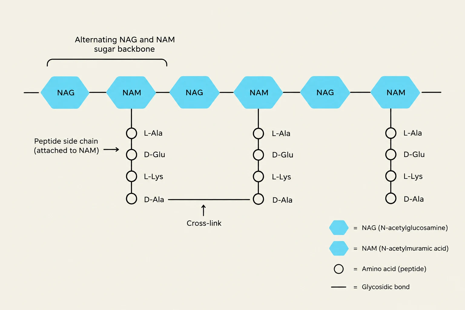
The peptidoglycan molecule is a mesh-like network composed of two main components: a glycan backbone and peptide cross-links. The glycan backbone consists of alternating sugar residues: N-acetylglucosamine (NAG) and N-acetylmuramic acid (NAM). These sugars form long, repeating carbohydrate strands that provide the basic scaffold of the wall.
To transform these individual strands into a rigid, load-bearing structure, short peptide side chains attached to the NAM residues must be connected to one another. These are known as peptide cross-links. This cross-linking turns the sugar chains into a continuous, stress-bearing network that provides the mechanical strength necessary to prevent the cell from bursting due to internal osmotic pressure.
This diagram illustrates the structure of peptidoglycan, a polymer consisting of alternating N-acetylglucosamine (NAG) and N-acetylmuramic acid (NAM) sugars, with peptide side chains attached to NAM and cross-links between these chains. This structure is a key component of bacterial cell walls.
Study Tip: Clinical Relevance of Peptidoglycan
Because peptidoglycan is unique to bacteria, drugs that target its synthesis are highly effective at killing bacteria without harming human host cells. However, remember that bacteria must constantly remodel this wall to grow, making actively dividing cells particularly vulnerable to these drugs.
Mechanisms of Disruption: Lysozyme and Beta-lactams
Two major biological and medical agents target different parts of this structure:
Lysozyme: A host enzyme (found in tears and saliva) that works by cleaving the chemical bond connecting NAG and NAM in the glycan backbone.
Beta-lactam antibiotics: These drugs (such as penicillin) interfere with the enzymes responsible for creating the peptide cross-links during new cell-wall synthesis.
In both cases, the result is a weakened wall that leaves the bacterium susceptible to osmotic rupture.
Gram-Positive vs. Gram-Negative Architecture
The way a bacterium organizes its peptidoglycan and membranes determines its "Gram status," which is revealed through Gram staining.
Gram-Positive Bacteria
Gram-positive bacteria are characterized by a relatively thick, robust layer of peptidoglycan surrounding a single cytoplasmic membrane. This thick wall provides significant mechanical strength. In many species, the wall is further reinforced by teichoic acids.
Wall teichoic acids are covalently bound to the peptidoglycan.
Lipoteichoic acids are anchored deeper, in the cytoplasmic membrane.
These negatively charged polymers help organize the envelope, influence surface charge, and assist in interacting with ions and host tissues.
Gram-Negative Bacteria
Gram-negative bacteria possess a much more complex, multi-layered envelope. Instead of one thick wall, they have a thin layer of peptidoglycan located within a specialized compartment called the periplasm. This periplasm sits between the inner cytoplasmic membrane and an additional outer membrane.
The outer membrane acts as a sophisticated permeability barrier, restricting the entry of many potentially harmful molecules, such as certain antibiotics and detergents. To allow necessary nutrients to pass through this barrier, Gram-negative bacteria utilize porins—channel-forming proteins that provide controlled pathways for small hydrophilic molecules.
Diagram illustrating the structural differences between Gram-positive and Gram-negative bacteria, highlighting their cell walls and membranes.
The Lipopolysaccharide (LPS) Layer
A defining feature of the Gram-negative outer membrane is the presence of Lipopolysaccharide (LPS) in its outer leaflet. LPS is not a uniform substance but is composed of three distinct structural regions:
Lipid A: This region anchors the LPS molecule into the outer membrane. Crucially, Lipid A is the "endotoxic" portion of the molecule; when released during cell lysis, it can trigger intense inflammatory responses in a host.
Core Polysaccharide: A central region connecting Lipid A to the O-antigen.
O-antigen: The outward-facing polysaccharide chain. Because the sugar composition of the O-antigen varies widely between different strains, it is often used for serotyping (distinguishing between different versions of the same bacteria).
Exceptions to the Rule: Acid-Fast and Wall-less Bacteria
Not all bacteria follow the standard Gram-positive/Gram-negative dichotomy.
Acid-Fast Bacteria:
Species like Mycobacteria possess a unique, lipid-rich envelope. Their peptidoglycan is linked to arabinogalactan and covered by a thick layer of long-chain mycolic acids. This high lipid content creates an extremely low-permeability barrier that slows the movement of many chemicals and provides high chemical resilience. Because of this, they do not stain well with standard Gram stains and require specialized "acid-fast" staining techniques.
Wall-less Bacteria (Mycoplasma):
Some bacteria, such as Mycoplasma species, lack a peptidoglycan cell wall entirely. They rely on a strengthened plasma membrane containing sterols for stability. Because they lack a target, beta-lactam antibiotics are intrinsically ineffective against them.
Summary of Envelope Diversity
While the bacterial cell envelope is a common theme, it is not universal across all prokaryotes. Archaea, for instance, use fundamentally different chemistry. They do not make peptidoglycan; instead, they may use pseudopeptidoglycan, protein S-layers, or polysaccharides. Their membranes also utilize ether-linked isoprenoid chains rather than the ester-linked fatty acids found in bacteria.
Understanding these structural nuances is clinically vital. The envelope architecture dictates whether a drug can enter the cell, how the immune system perceives the invader, and how the bacterium survives in hostile environments.
Diagrams illustrating the three major types of prokaryotic cell envelopes: Gram-positive, Gram-negative, and Acid-fast, detailing their structural components like peptidoglycan, outer membrane, LPS, porins, and mycolic acids.
Keep reading for free.
Create an account to finish this reading and make it part of your study routine.