Understanding how the body maintains internal stability requires a deep dive into the architecture of the nervous system. While we often think of our movements as conscious decisions, a vast network of involuntary signals is constantly working behind the scenes to regulate our heart rate, digestion, and respiratory function. This regulation is the domain of the autonomic nervous system (ANS), a specialized division of the peripheral nervous system designed to manage the "automatic" functions of our visceral organs.
The Architecture of the Nervous System
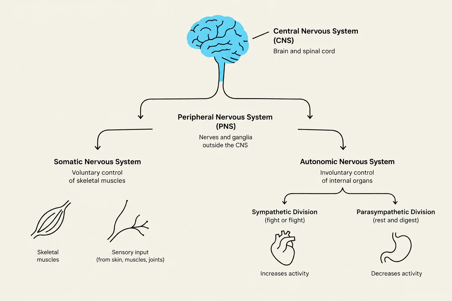
The human nervous system is organized into two primary branches: the Central Nervous System (CNS) and the Peripheral Nervous System (PNS). The CNS serves as the command center, consisting of the brain and the spinal cord, where integration and control occur. In contrast, the PNS acts as the communication highway, consisting of cranial and spinal nerves that connect the CNS to the rest of the body.
The PNS is further subdivided into two functional pathways:
The Somatic Division: This handles voluntary control. It conducts sensory (afferent) impulses from the body to the CNS and sends motor (efferent) impulses from the CNS to the skeletal muscles.
The Autonomic Nervous System (ANS): This handles involuntary control. It conducts visceral motor impulses from the CNS to cardiac muscle, smooth muscle, and glands.
Diagram illustrating the divisions of the human nervous system, including the Central Nervous System (CNS) and Peripheral Nervous System (PNS), with further breakdown into Somatic and Autonomic Nervous Systems, and their respective functions.
The Two-Neuron Chain
Unlike the direct pathways found in the somatic nervous system, autonomic pathways utilize a unique "two-neuron chain" to reach their target tissues. This chain consists of two distinct neurons that meet at a junction called a ganglion (a collection of nerve cell bodies located in the PNS).
1. The Preganglionic Neuron
The first neuron in the chain is the preganglionic neuron. Its cell body is housed within the CNS—either in the brainstem or the intermediolateral (IML) gray matter of the spinal cord. The axon of this neuron is myelinated, allowing for efficient signal transmission from the CNS to the ganglion.
2. The Postganglionic Neuron
The second neuron is the postganglionic neuron. Its cell body is located within a ganglion in the PNS. The axon of this neuron is unmyelinated and projects from the ganglion directly to the target tissue, such as a gland or a smooth muscle layer.
Exam Tip: Pay close attention to the relative lengths of these axons. In the Sympathetic Nervous System, preganglionic axons are typically short, while postganglionic axons are long. In the Parasympathetic Nervous System, the pattern is reversed: preganglionic axons are long, and postganglionic axons are short.
Sympathetic vs. Parasympathetic Divisions
The ANS is functionally split into two opposing divisions that work in tandem to maintain homeostasis: the Sympathetic Division and the Parasympathetic Division.
The Sympathetic Division (Fight-or-Flight)
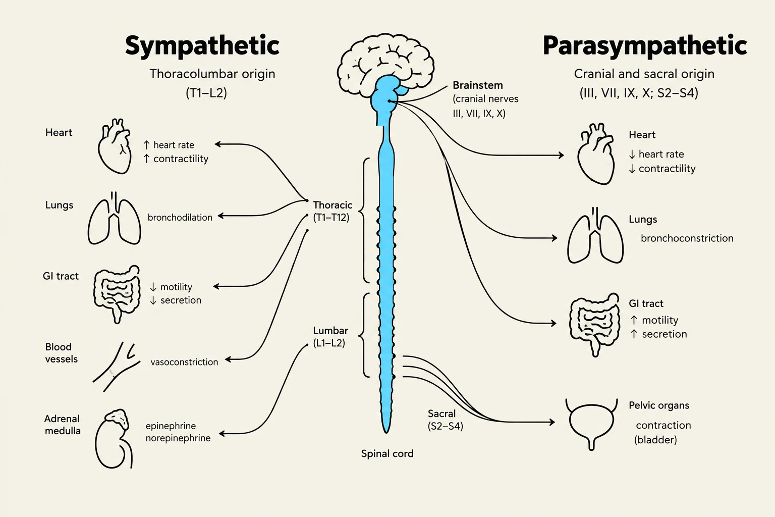
The sympathetic division prepares the body for intense physical activity or perceived threats. It is often referred to as the "thoracolumbar" outflow because its preganglionic neurons originate in the spinal cord from segments T1 through L2/L3. This system liberates neurotransmitters like epinephrine and norepinephrine to trigger widespread physiological changes.
The Parasympathetic Division (Rest-and-Digest)
The parasympathetic division promotes "housekeeping" functions, such as digestion and energy conservation. It is characterized by a "craniosacral" outflow, meaning its nerves originate from the cranial nerves (specifically CN III, VII, IX, and X) and the sacral segments of the spinal cord (S2–S4). The Vagus nerve (CN X) is particularly vital, serving as the primary preganglionic supply to the thoracic and abdominal viscera.
Diagram illustrating the sympathetic and parasympathetic nervous systems, their origins, and their effects on various organs such as the heart, lungs, GI tract, blood vessels, adrenal medulla, and pelvic organs.
Splanchnic Nerves and Autonomic Pathways
A key term in autonomic anatomy is the splanchnic nerve. A splanchnic nerve is defined as any nerve that carries autonomic (sympathetic or parasympathetic) fibers to the body cavity viscera.
In the sympathetic system, these nerves follow various routes. Some fibers synapse in the paravertebral sympathetic trunk (the columns of ganglia flanking the spine), while others pass through the trunk without synapsing to reach prevertebral ganglia located near the large blood vessels of the abdomen. These "thoracic splanchnic nerves" are essential for innervating organs like the liver, stomach, and kidneys.
Comparative Target Organ Effects
The true clinical significance of the ANS lies in how these two divisions produce opposite effects on the same organs to balance the body's needs.
Target Organ
Sympathetic Effect (Fight-or-Flight)
Parasympathetic Effect (Rest-and-Digest)
Eyes
Mydriasis (Pupil dilation) for maximum visual range
Miosis (Pupil constriction) to protect the retina
Lungs
Bronchodilation for maximum oxygen exchange
Bronchoconstriction and increased secretions
Heart
Increased heart rate (Chronotropy) and contractile force
Stimulates motility and enzyme secretion; relaxes internal anal sphincter
Skin/Glands
Piloerection, vasoconstriction, and sweating (sudomotor)
No effect on skin or sweat glands
Clinical Note: Recognizing these states is vital in a clinical setting. A patient in "sympathetic overdrive" may be in a state of high stress, which can make it difficult for them to learn new movements or exercise safely.
Sensory Feedback and Referred Pain
The autonomic system is not purely motor; it also has an afferent (sensory) function. Visceral afferent fibers transmit information about the internal environment—such as blood pH, heart rate, and organ distention—back to the CNS to help regulate homeostasis.
Because these visceral sensory fibers often travel alongside sympathetic fibers, the brain sometimes struggles to pinpoint the exact source of a signal. This results in referred pain, where pain from a visceral organ is felt in a different area (a specific dermatome) because the brain misinterprets the incoming signal.
Keep reading for free.
Create an account to finish this reading and make it part of your study routine.