Autonomic Nervous System: Motor Innervation and Divisions
Explains the autonomic nervous system's two-neuron pathway, its sympathetic ("fight-or-flight") and parasympathetic ("rest-and-digest") divisions, and the neurotransmitters involved in motor innervation.
The Autonomic Nervous System (ANS) is the regulatory powerhouse of the body, managing the involuntary functions required to maintain homeostasis. While the somatic nervous system governs our voluntary movements by communicating with skeletal muscles, the ANS provides motor innervation to visceral effectors, including smooth muscle, cardiac muscle, and glands. To achieve this, the ANS utilizes a specialized two-neuron pathway that allows for precise or widespread control depending on the body's needs.
The Two-Neuron Pathway
The diagram illustrates the two-neuron pathway of autonomic motor innervation, showing the connection from the central nervous system (CNS) through a preganglionic neuron, an autonomic ganglion, and an unmyelinated postganglionic neuron to a target organ like smooth muscle.
Unlike the direct connection seen in somatic motor neurons, autonomic motor innervation requires two successive neurons to travel from the Central Nervous System (CNS) to the target organ.
Preganglionic Neurons: These neurons have their cell bodies located within the CNS. They are characterized by being myelinated, which allows for relatively rapid signal conduction to the first synapse.
Postganglionic Neurons: These neurons originate in an autonomic ganglion (a cluster of nerve cell bodies located outside the CNS) and extend to the effector organ. These axons are unmyelinated, resulting in slower conduction speeds.
The synapse between these two neurons occurs within an autonomic ganglion, where neurotransmitters are released to pass the signal forward.
Functional Divisions: Sympathetic vs. Parasympathetic
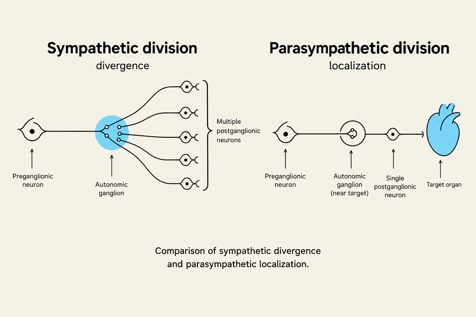
Comparison of sympathetic divergence and parasympathetic localization in the nervous system.
The ANS is divided into two functional components that typically exhibit dual innervation. This means that most body organs receive fibers from both divisions, which usually exert opposing effects to maintain a balance known as autonomic tone.
The Sympathetic Division ("Fight-or-Flight")
The sympathetic division is designed for physical or emotional stress, preparing the body for "Exercise, Excitement, Emergency, or Embarrassment". When activated, it triggers the "fight-or-flight" response, which includes dilating pupils, increasing heart rate and blood pressure, and diverting blood flow from the digestive tract to skeletal muscles.
Anatomy: Preganglionic cell bodies are located in the lateral horns of the thoracic and the first two or three lumbar segments of the spinal cord.
Ganglia and Pathways: Axons exit the spinal cord via white rami communicantes to enter the sympathetic trunk (paravertebral) ganglia. They may also pass through to prevertebral ganglia (such as the celiac or mesenteric ganglia) located near the abdominal organs.
Divergent Projection Pattern: A key feature of the sympathetic division is its high degree of divergence. A single preganglionic neuron often synapses with many postganglionic neurons. This allows for a widespread, simultaneous, and intense body-wide response.
The Adrenal Medulla: This serves as a specialized complex of postganglionic neurons. When stimulated, it releases epinephrine and norepinephrine directly into the bloodstream, which intensifies and prolongs the sympathetic response.
The Parasympathetic Division ("Rest-and-Digest")
The parasympathetic division conserves energy and promotes "rest-and-digest" activities, such as digestion and waste elimination. Its effects are often summarized by the acronym SLUDD: Salivation, Lacrimation, Urination, Digestion, and Defecation.
Anatomy: Preganglionic cell bodies are located in the brain stem (specifically cranial nerves III, VII, IX, and X) and the sacral segments (S2–S4) of the spinal cord.
Terminal Ganglia: Unlike the sympathetic division, parasympathetic neurons form terminal ganglia located very near or even within the walls of the target organ.
Localized Response: Because the parasympathetic division has a much lower degree of divergence, its effects can be highly localized to a specific organ without affecting the entire body simultaneously.
Exam Tip: Comparison of Divergence
If a question asks which division is better suited for a coordinated, whole-body response (like a sudden scare), choose Sympathetic due to its divergent projection pattern. If it asks which division allows for precise, organ-specific control (like slowing the heart without stopping digestion), choose Parasympathetic.
Neurotransmitters and Receptor Classification
Autonomic neurons are classified by the specific neurotransmitter they release at the synapse with the effector organ.
Cholinergic Neurons
These neurons release acetylcholine (ACh).
All preganglionic neurons (both sympathetic and parasympathetic) are cholinergic.
All parasympathetic postganglionic neurons are cholinergic.
Sympathetic postganglionic neurons that innervate sweat glands are also cholinergic.
Cholinergic receptors are divided into two types: Nicotinic (which always cause excitation/depolarization) and Muscarinic (which can cause either excitation or inhibition depending on the target organ).
Adrenergic Neurons
These neurons release norepinephrine (NE).
Most sympathetic postganglionic neurons are adrenergic.
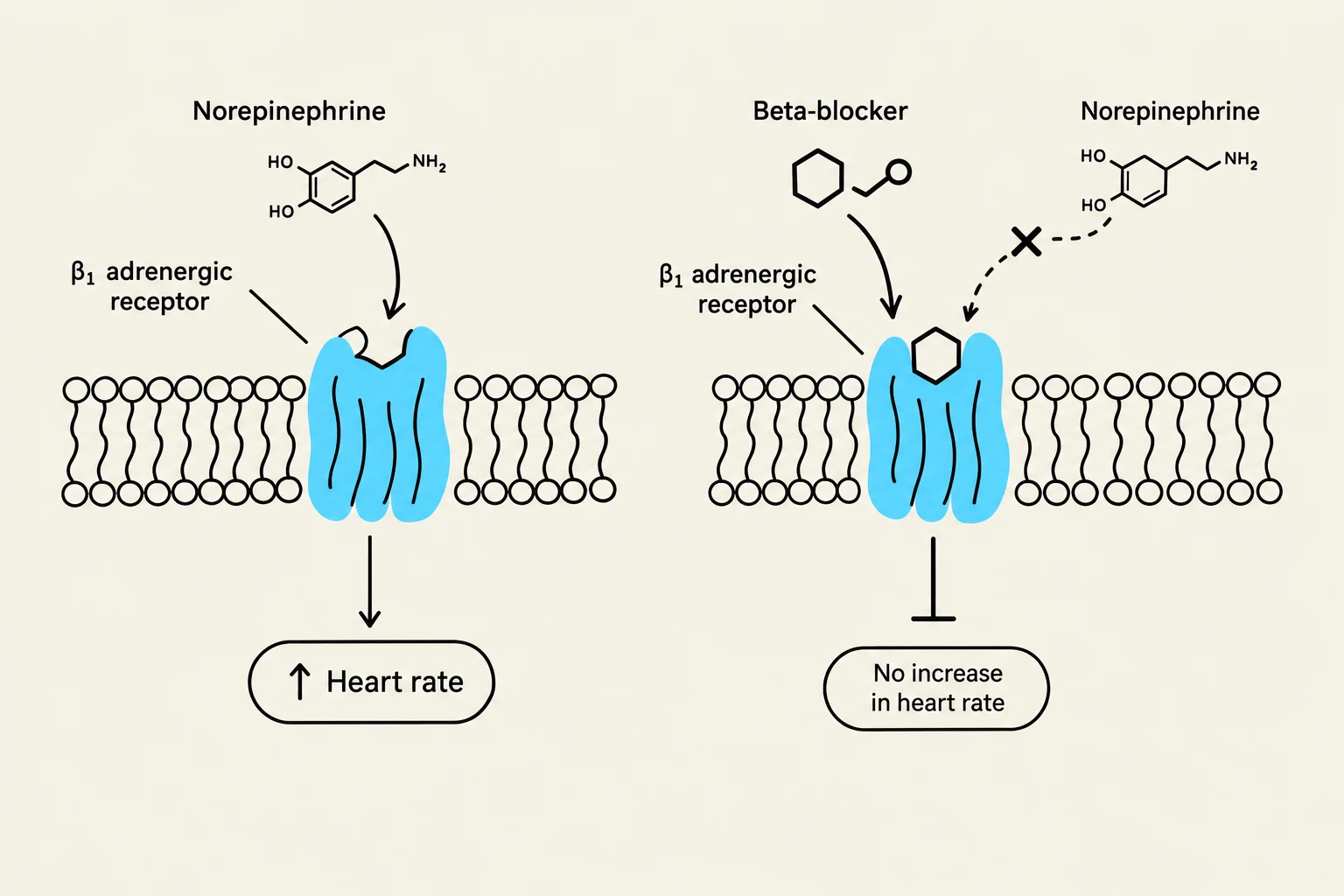
Adrenergic receptors are classified into α (alpha) and β (beta) subtypes. For example, β1 receptors are found predominantly in the heart, where their activation increases heart rate.
Summary Table of Neurotransmitters
Synapse
Sympathetic Division
Parasympathetic Division
First Synapse (Preganglionic)
ACh (Nicotinic)
ACh (Nicotinic)
Second Synapse (Postganglionic)
NE (Adrenergic)
ACh (Muscarinic)
Exception (Sweat Glands)
ACh (Muscarinic)
N/A
Clinical Correlations
Diagram illustrating the mechanism of action of norepinephrine and beta-blockers on the beta-1 adrenergic receptor, affecting heart rate.
Understanding the ANS is vital for understanding various medical conditions and pharmacological treatments:
Raynaud Disease: This condition involves excessive sympathetic stimulation of the smooth muscle in the arterioles of the fingers and toes, causing them to become ischemic (lacking blood) when exposed to cold or stress.
Beta Blockers: These are drugs used to treat cardiovascular issues. They attach to β1 receptors in the heart, blocking the effects of adrenaline and thereby slowing the heart rate and reducing arrhythmia.
Atropine: An anticholinergic drug that blocks parasympathetic effects. It is used to suppress secretions during surgery or to dilate the pupils during eye exams.
Depression Treatment: Since norepinephrine is often viewed as a "feel-good" neurotransmitter, drugs like MAO inhibitors (which prevent the inactivation of NE) or Tricyclic antidepressants are used to prolong the activity of NE in the synapse to help relieve depression.
Keep reading for free.
Create an account to finish this reading and make it part of your study routine.