An overview of autoimmune diseases, their underlying mechanisms, and the laboratory methods used for diagnosis, including antinuclear antibody (ANA) testing and various immunoassay techniques.
Autoimmune diseases represent a failure of the immune system to maintain self-tolerance, leading to an immune response directed against the body's own tissues. These conditions can be broadly categorized into systemic diseases, which affect multiple organ systems, and organ-specific diseases, which target a single tissue or cell type. Understanding the underlying immunopathogenic mechanisms and mastering the various laboratory methods used to detect autoantibodies is essential for clinical diagnosis and monitoring.
Autoimmune Disease Mechanisms
The fundamental mechanism of autoimmunity involves the production of autoantibodies—antibodies that target self-antigens. The interaction between an antibody (Ab) and an antigen (Ag) is governed by the law of mass action, where the binding is a dynamic and reversible process:
AGN+ABY⟶AGNABY complex=Keq=K1/K2
In this equation, K1 represents the rate of the forward reaction and K2 represents the rate of the reverse reaction. The stability of the complex depends on affinity (the initial force of attraction between a single Fab site and one epitope) and avidity (the sum of all antibody-epitope pairs).
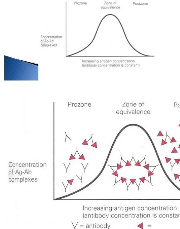
In many autoimmune processes, these interactions lead to the formation of immune complexes that trigger inflammation or tissue destruction. A critical concept in laboratory testing is the zone of equivalence. For visible reactions like agglutination or precipitation to occur, the ratio of antigen to antibody must be optimal:
Prozone: An excess of antibody prevents the formation of a lattice, potentially leading to false-negative results.
Zone of Equivalence: The optimal ratio where maximum binding and lattice formation occur.
Postzone: An excess of antigen prevents effective cross-linking.
This graph illustrates the relationship between antigen concentration and the concentration of antigen-antibody complexes, showing the prozone, zone of equivalence, and postzone.
Systemic and Connective Tissue Diseases
Systemic autoimmune diseases affect various parts of the body simultaneously. A primary example is Systemic Lupus Erythematosus (SLE). The pathogenesis of SLE is complex, often involving the production of antibodies against nuclear components like DNA and histones.
Systemic Lupus Erythematosus (SLE)
SLE is characterized by a wide array of autoantibodies. Specific markers include:
Anti-Smith (anti-Sm) antibodies: Highly specific for SLE.
Anti-double stranded DNA (dsDNA): Highly specific for SLE; the presence of these can be confirmed using the Crithidia test, which uses Crithidia luciliae cells to detect native DNA.
Diagram of a Trypanosome, showing the nucleus, kinetoplast, and flagella.
Connective Tissue Diseases
Other systemic conditions include Sjögren Syndrome, Mixed Connective Tissue Disease (MCTD), and Progressive Systemic Sclerosis (Scleroderma). In Scleroderma, the presence of Scl-70 antibodies is a marker for a poor prognosis, whereas centromere antibodies are associated with the CREST syndrome and a better prognosis.
Rheumatoid Arthritis (RA)
Rheumatoid arthritis is a chronic inflammatory disorder. Immunologic findings often include:
Rheumatoid Factor (RF): An IgM antibody directed against the Fc portion of IgG.
Anti-CCP (Cyclic Citrullinated Peptide): A highly specific test for RA.
Associated markers: Elevated C-reactive protein (CRP), increased Erythrocyte Sedimentation Rate (ESR), and associations with HLA-DR4 alleles.
Organ-Specific Autoimmune Diseases
Unlike systemic diseases, organ-specific diseases target a particular functional unit of the body. Examples include:
Hashimoto's Thyroiditis: Associated with HLA-DR3 and DR4.
Myasthenia Gravis: Associated with HLA-DR3.
Addison's Disease: Associated with HLA-DR3.
Graves' Disease: Associated with HLA-B08, C07, DR3, and DR7.
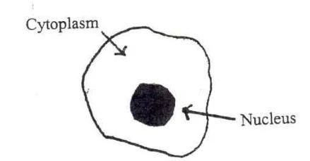
The ANA test is a primary screening tool for many autoimmune diseases. It is typically performed using Indirect Immunofluorescence (IFA) or ELISA. When using IFA, the pattern of fluorescence in the cell nucleus provides vital diagnostic clues.
Diagram of a cell showing the cytoplasm and nucleus.
ANA Patterns and Antigens
The following table summarizes common patterns observed during ANA testing:
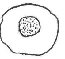
A simple diagram of a cell nucleus.A simple diagram of a cell with a nucleus.
Exam Tip: The Speckled Pattern
To distinguish the speckled pattern from nucleolar or centromere patterns, look for "spots of no staining" within the nucleus.
Advanced Laboratory Methods
Agglutination and Precipitation
Direct Agglutination: Detects antigens naturally present on particles (e.g., bacteria or RBCs).
Passive Agglutination: Uses carrier particles like latex beads coated with known antigens to detect unknown antibodies.
Reverse Passive Agglutination: Uses particles coated with known antibodies to detect unknown antigens.
Precipitation: Involves soluble antigens and antibodies forming insoluble complexes. Methods include Ouchterlony Double Diffusion and Rocket Immunoelectrophoresis.
Labeled Immunoassays (ELISA, RIA, and Western Blot)
For antigens or antibodies present in very low concentrations, labeled assays are required:
Radioimmunoassay (RIA): Uses radioisotopes (like 125I) as labels. It is highly sensitive but carries radiation hazards.
Enzyme Immunoassay (EIA/ELISA): Uses enzymes to produce a colorimetric, fluorescent, or luminescent signal. It is safer and more common than RIA.
Sandwich ELISA: "Sandwiches" the antigen between a capture antibody and a detection antibody.
Indirect ELISA: Typically used to detect antibodies by using a secondary enzyme-labeled anti-human antibody.
Western Blot: An adaptation of immunofixation electrophoresis where proteins are transferred to a nitrocellulose membrane. It is used for complex antigenic compositions, such as confirming HIV antibodies.
Molecular Methods
Polymerase Chain Reaction (PCR) is used to amplify specific DNA sequences, allowing for the detection of pathogens or genetic markers that are difficult to culture.
Clinical Significance of Titers
When monitoring a patient, a four-fold rise in antibody titer (an increase of two doubling dilutions, e.g., from 1:4 to 1:16) is considered clinically significant.
Keep reading for free.
Create an account to finish this reading and make it part of your study routine.