Explains the pathophysiology of asthma, including airway inflammation, bronchoconstriction, and mucus production. Details pharmacological management with rescue and controller medications, and discusses clinical red flags and emergencies like status asthmaticus.
Asthma is a chronic respiratory condition defined by reversible airway obstruction. Unlike some other chronic lung diseases where the damage is permanent, asthma is characterized by episodes of narrowing that can be reversed, though the underlying driver is a persistent state of airway inflammation. Understanding asthma requires looking beyond the simple sound of a wheeze; it requires an understanding of the complex immune responses and physiological changes that occur within the lungs.
The Pathophysiology of Asthma
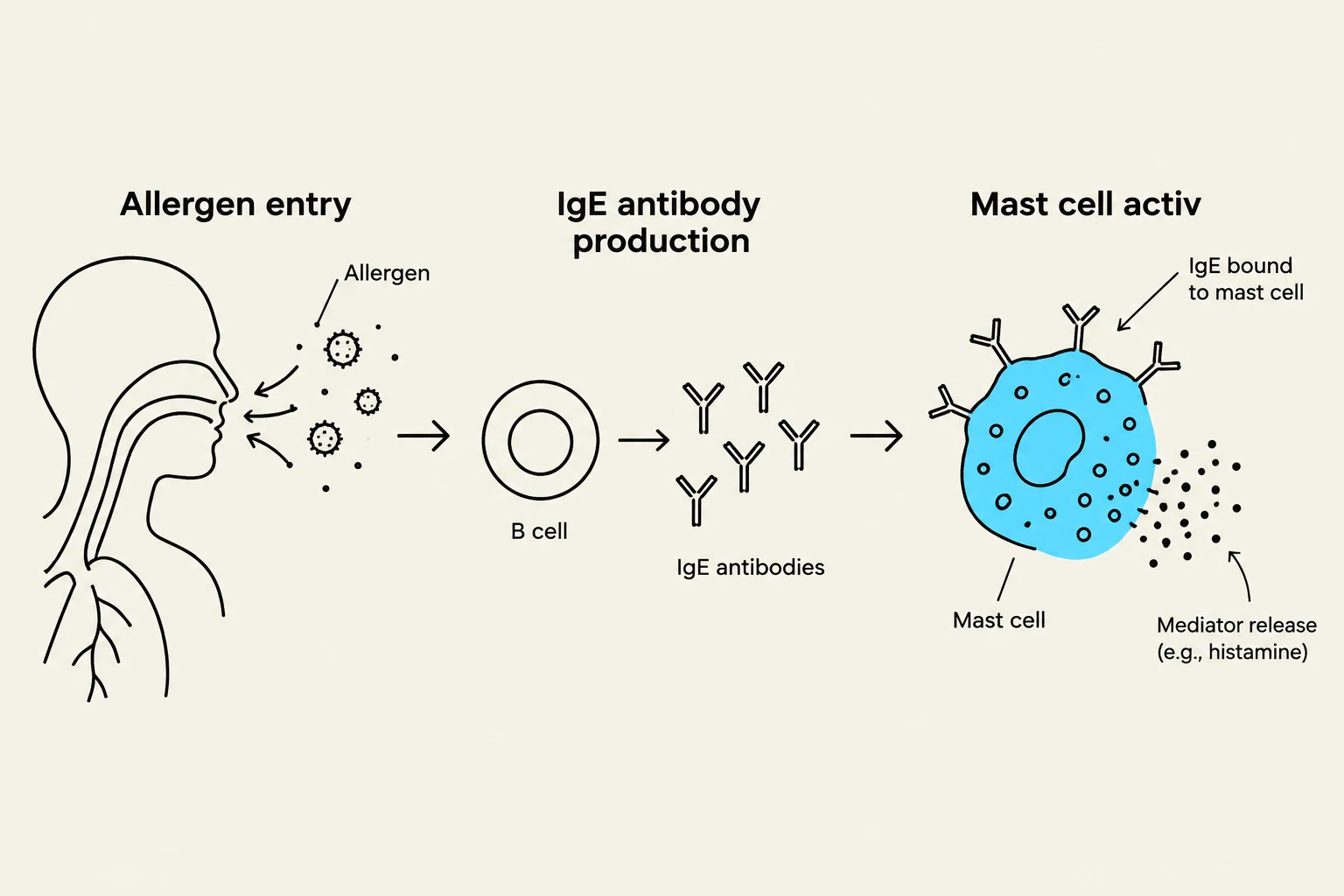
At its core, asthma is an inflammatory disease. While many patients experience "allergic asthma," the process begins when an allergen—such as pollen, dust mites, or pet dander—enters the airway. This triggers an immune overreaction involving a specific cascade of biological actors:
IgE Antibodies: The immune system produces Immunoglobulin E (IgE) in response to the perceived threat.
Mast Cell Activation: These IgE antibodies bind to mast cells located in the airway tissues.
Chemical Release: Once activated, mast cells release potent inflammatory mediators, including histamine, leukotrienes, and prostaglandins.
These chemicals act on the surrounding tissues to initiate the physical symptoms of an attack.
Diagram illustrating the process of an allergic reaction, from allergen entry into the respiratory system, to IgE antibody production by B cells, and finally, mast cell activation leading to mediator release.
The "Three S's" of Airway Obstruction
The release of inflammatory chemicals leads to three distinct physiological changes that collectively narrow the airway diameter:
Swelling: The airway lining becomes inflamed and edematous (swollen).
Spasm: The smooth muscles surrounding the airways tighten, a process known as bronchoconstriction.
Secretions: The body produces excess mucus, which clogs the already narrowed passages.
These changes manifest clinically as the classic symptoms of asthma: wheezing, coughing, chest tightness, and shortness of breath.
Health Disparities and Environmental Factors
Asthma is not distributed equally across all populations. Socioeconomic factors and historical systemic issues play a significant role in asthma prevalence and severity.
A primary driver of these disparities is redlining—a historical practice that has led to long-term socioeconomic disadvantages in specific neighborhoods. Residents in these areas often face higher asthma rates due to:
Poor Housing Quality: Increased exposure to mold, pests, and indoor allergens.
Environmental Pollution: Higher proximity to highways, industrial zones, and heavy traffic.
Reduced Access: Limited availability of healthcare and community resources to manage the condition.
Pharmacological Management
Effective asthma management requires a dual approach: treating the immediate symptoms and addressing the underlying chronic inflammation.
1. Rescue Medications (The "Openers")
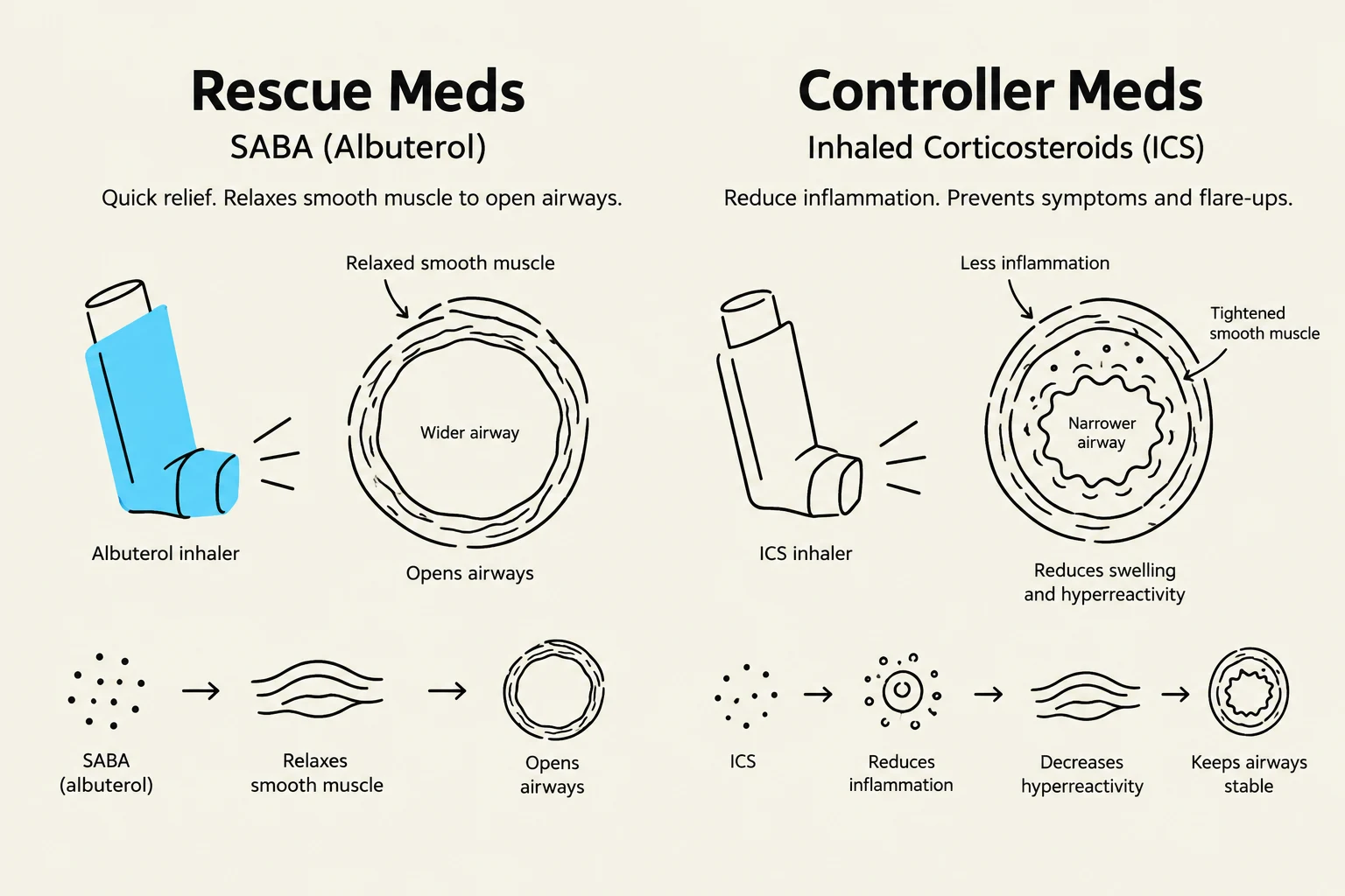
When a patient experiences an acute attack, they require rescue medication to provide rapid relief. These are typically Short-Acting Beta-Agonists (SABA), such as albuterol.
Mechanism: They work by relaxing the bronchial smooth muscle.
Goal: To "open the airway" by reversing bronchoconstriction quickly.
2. Controller Medications (The "Calmers")
To prevent attacks from occurring in the first place, patients use controller medications. The most common type is inhaled corticosteroids.
Mechanism: They target the underlying inflammatory process.
Goal: To "calm the airway" by reducing chronic swelling and sensitivity over time.
This diagram illustrates the mechanisms of rescue medications (SABA like Albuterol) and controller medications (ICS) for managing asthma. Rescue inhalers provide quick relief by relaxing smooth muscles to widen airways. Controller inhalers reduce inflammation, preventing symptoms and flare-ups by decreasing swelling and hyperreactivity, thus keeping airways stable.
Clinical Red Flags and Emergencies
As a healthcare provider or student, recognizing the progression from a mild attack to a life-threatening emergency is critical.
The Danger of the "Silent Chest"
In a severe asthma attack, a patient may exhibit accessory muscle use, restlessness, and an inability to speak in full sentences. However, a sudden decrease in wheezing can be a major red flag. This is known as a "silent chest," which indicates that there is so little airflow moving through the lungs that a wheezing sound can no longer be produced. This suggests imminent respiratory failure.
Interpreting Arterial Blood Gases (ABGs)
ABGs are vital tools for monitoring the severity of an exacerbation. The progression typically follows this pattern:
Early Stage (Respiratory Alkalosis): The patient hyperventilates to compensate for the obstruction. This results in low CO2 levels and a high pH.
Worsening Stage (The Red Flag): If a distressed patient presents with a normalPaCO2, it indicates they are tiring out and can no longer maintain the hyperventilation required to compensate.
Late Stage (Respiratory Acidosis): As the patient reaches respiratory failure, they can no longer exhale CO2, leading to CO2 retention, a low pH, and severe hypoxemia.
Exam Tip: The CO2 Trend
In an asthma attack, a risingPaCO2 is often much more concerning than a low one. A low CO2 means the patient is still fighting; a normal or high CO2 means the patient is exhausting their respiratory muscles and heading toward failure.
Status Asthmaticus
When an asthma attack is severe and unresponsive to routine treatments, it is classified as Status Asthmaticus. This is a medical emergency that can rapidly progress to respiratory failure and requires immediate intensive intervention.
Keep reading for free.
Create an account to finish this reading and make it part of your study routine.