Assessment of the Breasts and Regional Lymphatic System
A clinical guide to assessing the breasts and regional lymphatic system, covering developmental changes, lactation-related disorders, and physical examination techniques for identifying potential malignancies.
The assessment of the breasts and the regional lymphatic system is a critical component of female reproductive health and general oncology screening. Because breast cancer remains the most common cancer in females, clinicians must possess a high degree of proficiency in distinguishing normal developmental variations from pathological findings. This guide explores the physiological changes across the lifespan, common lactation-related disorders, and the systematic physical examination techniques required to identify potential malignancies.
Developmental and Physiological Considerations
Breast tissue is highly dynamic, responding significantly to hormonal shifts throughout a person's life.
Infants and Adolescents
In infants, it is not uncommon to observe the secretion of clear or white fluid from the nipples. Adolescent development typically commences between the ages of 8 and 10; however, clinicians should be aware of precocious puberty, which is defined as the onset of secondary sex characteristics at age 8 or younger.
Pregnancy and Lactation
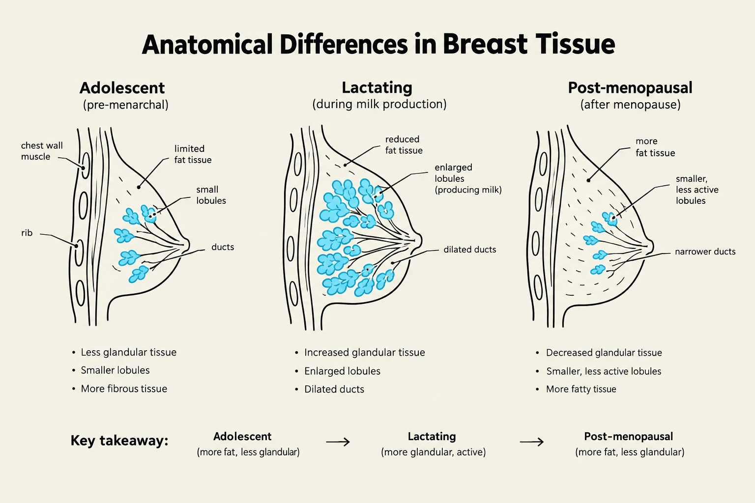
During pregnancy, the breasts undergo significant physiological changes to prepare for milk production. These include increased vascular patterns, changes in nipple appearance, and the production of colostrum (the first milk), which typically begins around four months of gestation. Individuals who are lactating may experience engorgement, redness, and nipple soreness.
Older Females and Males
Post-menopause, the decline in estrogen and progesterone levels leads to glandular atrophy and a loss of subcutaneous fat. This reduction in breast size and elasticity makes the inner structures and lactiferous ducts more palpable during examination.
In males, the anatomy typically consists of a small disc of undeveloped tissue underlying the nipple. A common clinical finding in males is gynecomastia, which refers to the temporary enlargement of breast tissue, often occurring during puberty.
This diagram illustrates the anatomical differences in breast tissue across three life stages: adolescent (pre-menarchal), lactating (during milk production), and post-menopausal (after menopause), highlighting changes in glandular tissue, fat tissue, lobules, and ducts.
Lactation-Related Disorders
When breastfeeding or chestfeeding, several localized disorders may arise due to milk stasis or infection.
Plugged Duct: This occurs when a single milk duct becomes clogged. It presents as a localized area of tenderness and may appear reddened, but it is not an infection. Management involves frequent emptying of the breast through feeding or expression.
Mastitis: This is an inflammatory infection that often occurs in a single quadrant. The area is typically red, swollen, hard, and very hot to the touch. Patients often present with systemic flu-like symptoms, including fever, chills, malaise, and increased pulse. Treatment requires rest, local heat, antibiotics, and continued frequent feeding.
Breast Abscess: A serious complication of mastitis where a pocket of pus accumulates in a localized area. If an abscess is suspected, nursing on the affected side must be temporarily discontinued; milk should be manually expressed and discarded. Treatment may involve antibiotics and surgical incision and drainage.
Clinical Assessment Techniques
A thorough assessment requires both subjective data collection (the patient's history) and objective data collection (the physical exam).
Subjective Data and Risk Factors
When taking a history, clinicians should ask about pain, lumps, discharge, rashes, and swelling. It is vital to identify both modifiable and non-modifiable risk factors for breast cancer.
Exam Tip: Identifying Urgent Findings
Always prioritize immediate follow-up if a patient reports unexplained nipple bleeding or discharge, presents with an open or ulcerating non-traumatic lesion, or if a new mass is discovered in a patient with a history of breast cancer.
Risk Factor Summary:
Non-modifiable: Female sex, age (50–69), personal/family history, BRCA1/BRCA2 mutations, Ashkenazi Jewish ancestry, and early menarche (before age 11) or late menopause (after age 55).
The physical exam should begin with inspection and proceed to palpation.
1. Inspection:
Observe the breasts for symmetry, size, and shape. Check the skin for color changes, redness, dimpling, edema, or rashes. Inspect the nipples for protrusion, retraction, scaling, fissures, or discharge.
2. Palpation:
Palpation should be performed systematically. For lumps, note the location, size, shape, consistency, movability, and distinctness. If discharge is present, note its color, consistency, and whether blood is present.
3. Lymphatic Assessment:
The regional lymphatic system includes the axilla (underarm) and supraclavicular regions. During the exam, support the patient's arm and palpate for tenderness, swelling, or palpable lymph nodes.
Diagram illustrating the breast quadrant method for palpation and axillary lymph node assessment.
Identifying Malignancy
While many breast changes are benign, certain characteristics are highly suspicious for breast cancer.
Characteristics of Breast Cancer
A malignant mass typically presents with the following "red flag" features:
Solitary and Unilateral: Usually found in only one breast.
Nontender: Unlike mastitis, cancerous masses are often painless.
Hard and Fixed: The mass feels dense and may be stuck to the underlying tissue or the skin.
Irregular Borders: The edges are poorly defined and not smooth.
Location: Most commonly found in the upper outer quadrant.
Advanced stages may manifest as skin dimpling, nipple retraction, or firm, hard axillary lymph nodes.
Breast Self-Examination (BSE)
Patients should be encouraged to perform regular breast self-examinations to monitor for any new lumps, redness, or swelling. Early detection through regular screening, such as mammography, is the most effective way to improve survival rates.
This illustration compares benign lumps and malignant masses, highlighting characteristics such as border regularity, mobility, and tenderness, and notes the involvement of axillary lymph nodes in malignant cases.
Keep reading for free.
Create an account to finish this reading and make it part of your study routine.