Apicomplexan Parasites: Cystoisospora, Toxoplasma, and Neospora
An overview of Apicomplexa, focusing on the genera Cystoisospora, Toxoplasma, and Neospora. It details their morphology, life cycles, transmission, clinical significance in various hosts, and general treatment principles.
The phylum Apicomplexa contains a diverse group of protists known as "coccidians". These parasites are characterized by complex life cycles that often involve multiple hosts and various developmental stages within the host's tissues. This article explores three significant genera within the order Eimeriorina: Cystoisospora, Toxoplasma, and Neospora.
Understanding Apicomplexa and Oocyst Morphology
Apicomplexans are part of the SAR supergroup of protists. A critical skill in veterinary parasitology is the ability to identify these organisms via fecal flotation by examining their oocysts—the thick-walled, resistant stages passed in feces.
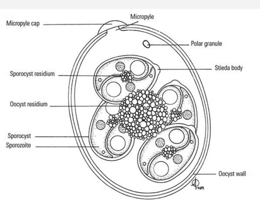
Distinguishing between different genera often relies on the internal structure of the oocyst. For example, a key diagnostic difference exists between Eimeria and Cystoisospora:
Eimeria spp.: Typically contain four sporocysts per oocyst.
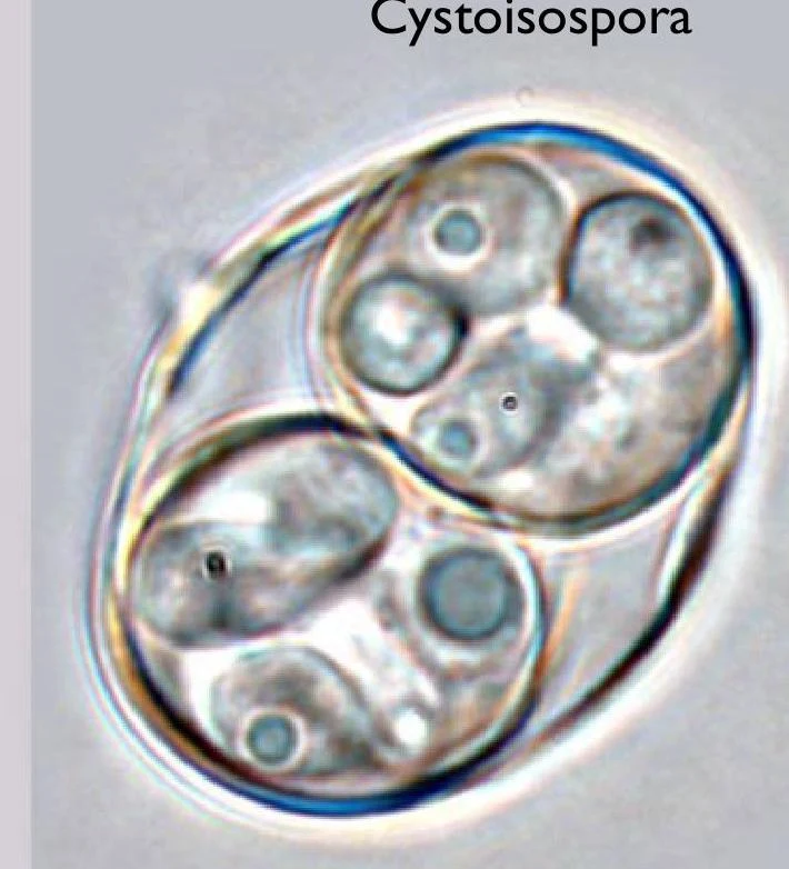
Cystoisospora spp.: Contain two sporocysts, each containing four sporozoites.
Diagram illustrating the structure of a coccidian oocyst, showing the micropyle, sporocysts, sporozoites, and residual bodies.
Cystoisospora is a genus of parasitic protozoa that cause coccidiosis.
Exam Tip: When performing a fecal flotation, always count the number of sporocysts within the oocyst to differentiate between Eimeria (4 sporocysts) and Cystoisospora (2 sporocysts).
Cystoisospora spp.
Cystoisospora species are enteric coccidians that primarily affect carnivores, such as canids (dogs) and felids (cats).
Life Cycle and Transmission
Infection occurs through the ingestion of sporulated oocysts from the environment or by consuming paratenic hosts (such as rodents, rabbits, or birds) that harbor the parasite. Once ingested, the parasite undergoes sexual stages within the carnivore host. The oocysts are then passed in the feces, where they must sporulate in the environment to become infectious.
Clinical Significance
Dogs and Cats: Can cause diarrhea, which may be hemorrhagic or nonhemorrhagic. In puppies and newborn kittens, this can lead to significant disease. Severe bloody diarrhea can result in anemia.
Pigs:Cystoisospora suis causes neonatal coccidiosis in 1–2 week old pigs, characterized by diarrhea, dehydration, and weight loss.
Treatment: Sulfadimethoxine and Triazines (such as ponazuril or toltrazuril) are used for treatment, though resistance is developing in some species.
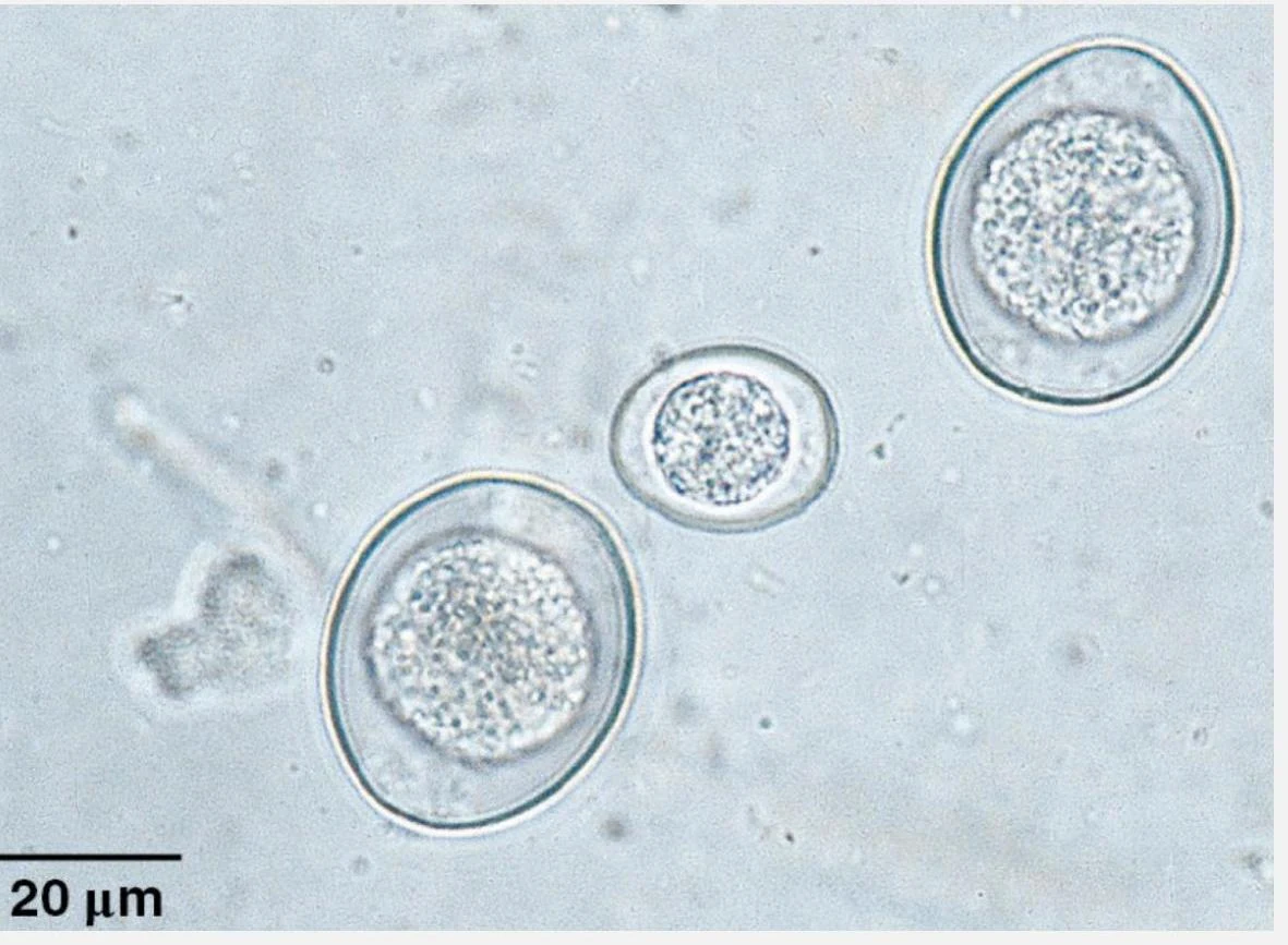
Microscopic view of parasitic ova, likely from a fecal sample, showing their characteristic oval shape and internal granular structures. The scale bar indicates 20 micrometers.
Toxoplasma gondii
Toxoplasma gondii is a highly significant enteric coccidian of felids. While many animals can serve as intermediate hosts, felids are the only definitive hosts capable of shedding oocysts.
The Complex Life Cycle
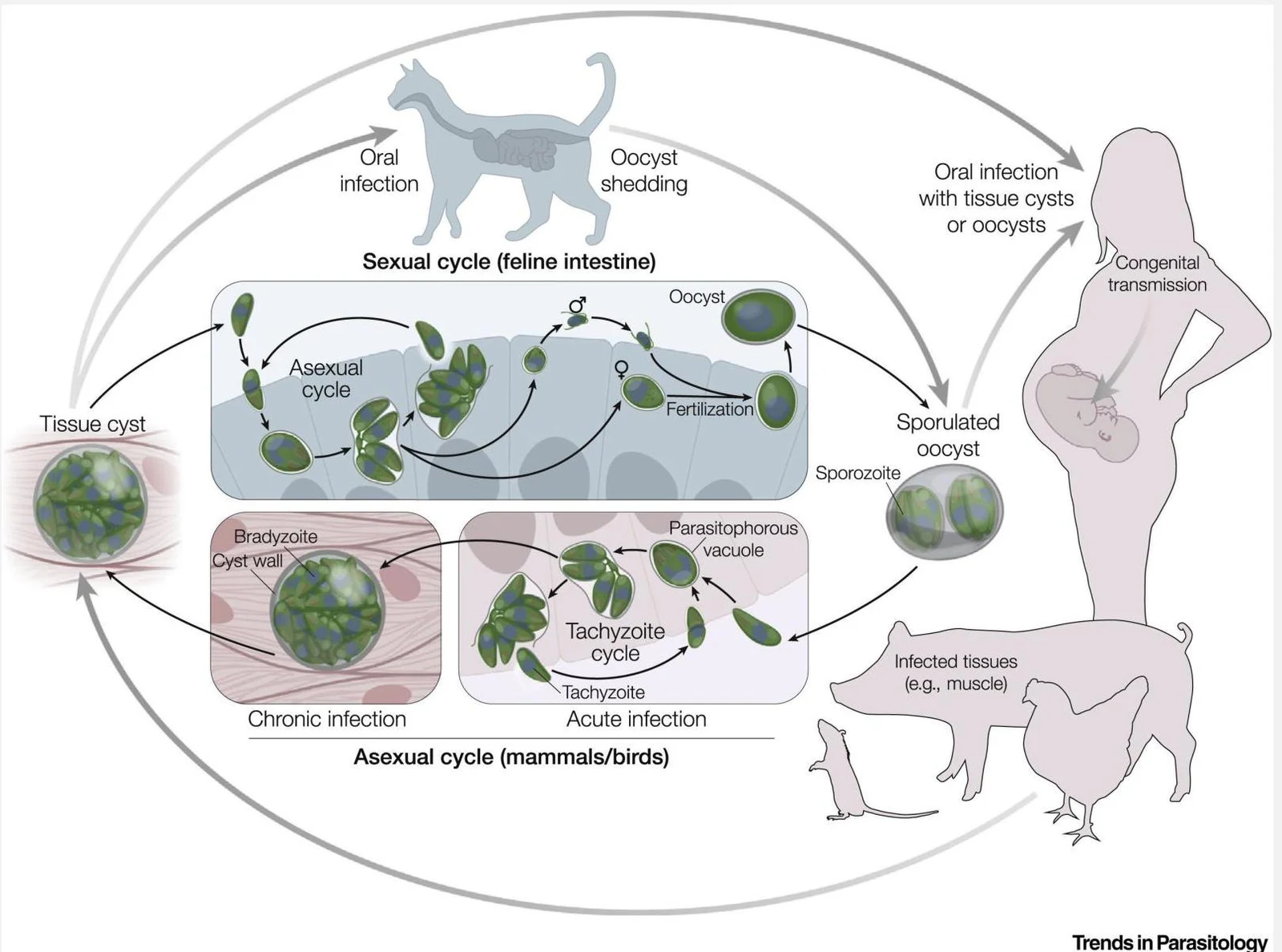
The life cycle involves a transition between different stages: sporozoites → tachyzoites → bradyzoites.
Definitive Host (Felid): An unexposed cat ingests bradyzoites in tissue cysts. The cyst ruptures, and bradyzoites infect epithelial cells, undergoing schizogony to form merozoites. These form gametes, resulting in the release of unsporulated oocysts in the feces.
Environment: Unsporulated oocysts sporulate within 1–5 days.
Intermediate Hosts: Any warm-blooded animal (including humans) can ingest sporulated oocysts. In these hosts, the parasite develops into tachyzoites (which cause rapid tissue destruction) and eventually into inactive bradyzoite cysts in muscle and neural tissues.
This diagram illustrates the life cycle of Toxoplasma gondii, a parasite that can infect various mammals and birds, including humans. It shows the sexual cycle in the feline intestine and the asexual cycle in intermediate hosts, leading to potential congenital transmission in humans.
Zoonotic Potential and Clinical Signs
Toxoplasma has massive zoonotic potential, affecting numerous wildlife species and humans. In humans, it can cause neurologic disease, ocular disease, and birth defects.
In cats, clinical disease is uncommon in immunocompetent individuals, but tachyzoites can cause granulomatous inflammation in organs like the liver, lungs, and brain. In immunocompromised cats, reactivated tissue cysts can cause disease.
Prevention for Humans (CDC Recommendations):
Cook meat to safe internal temperatures and freeze meat at sub-zero temperatures before cooking.
Rinse fruits and vegetables thoroughly.
Avoid unpasteurized goat's milk and raw shellfish.
If pregnant or immunocompromised, avoid changing cat litter and keep cats indoors to prevent hunting.
Neospora caninum
Neospora caninum is another important coccidian, primarily known for its impact on the livestock industry.
Life Cycle and Transmission
Definitive Host: Canids (dogs) shed oocysts in their feces.
Intermediate Host: Cattle are the primary intermediate hosts. They can be infected by ingesting oocysts from the environment or via transplacental infection from a carrier dam.
Clinical Disease: Neosporosis
In Cattle: It is a major cause of bovine abortion. This can present as endemic abortion (low-level, caused by reactivated bradyzoites in carrier cows) or epidemic abortion (large-scale "storms" caused by contaminated feed).
In Dogs: Puppies can suffer from transplacental infection, leading to progressive paralysis, myositis, or rigid hyperextension of the limbs. Adult dogs may develop neurologic signs if they become immunosuppressed.
Summary of Treatment and Pharmacology
While there are no approved treatments for Toxoplasma or Neospora in many species, certain drugs are used off-label in dogs.
Sulfonamide Drugs (e.g., Sulfadimethoxine, Sulfadiazine, Trimethoprim-sulfamethoxazole) work by inhibiting the synthesis of folic acid, which is essential for DNA and RNA synthesis. These drugs are primarily effective against the schizont (meront) stage of the parasite.
Clinical Note: When administering sulfonamides, ensure the patient remains adequately hydrated to prevent the formation of crystals in the urine (crystalluria).
Quick Review Table
Parasite
Primary Definitive Host
Key Clinical Sign
Diagnostic Feature
Cystoisospora
Canids/Felids
Enteric diarrhea
2 sporocysts per oocyst
Toxoplasma
Felids
Zoonotic; tissue cysts
Oocysts shed only by felids
Neospora
Canids
Bovine abortion
Transplacental transmission
Keep reading for free.
Create an account to finish this reading and make it part of your study routine.