This content explains anemia as a clinical finding, detailing its classification based on red blood cell morphology (size and color) and pathophysiology (regenerative vs. non-regenerative). It covers key metrics like MCV and MCHC, common causes of different anemia types, and the mechanisms of hemolysis.
Anemia is not a disease in itself, but rather a clinical or laboratory finding that reflects an underlying disease process. It is characterized by a decrease in red blood cell (RBC) count, hematocrit (HCT), and hemoglobin (Hgb), which ultimately results in a decreased ability of the blood to meet the oxygen demands of the animal. Patients may present with clinical signs such as pale mucous membranes, lethargy, weakness, increased heart rate, or even heart murmurs.
Depending on the speed of onset, anemia can be acute (such as severe blood loss leading to shock) or chronic (where the body has had time to physiologically adjust to lower oxygen levels). To understand an anemic patient, clinicians must classify the anemia using two primary methods: RBC morphology and pathophysiology.
RBC Morphology: Size and Color
The first way to classify anemia is by looking at the appearance of the red blood cells on a blood film. This involves assessing two main characteristics: size and color.
RBC Size and the MCV
The size of the RBCs is determined by the Mean Corpuscular Volume (MCV), which represents the average volume of all the RBCs in a sample. To calculate the MCV in a laboratory setting, you use the following formula:
MCV=RBCHct
Based on the MCV, we categorize the anemia into three types:
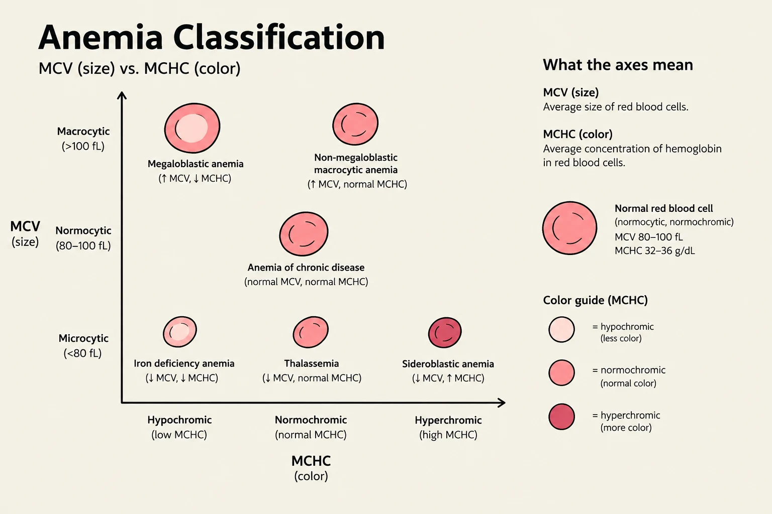
Macrocytic anemia: Increased MCV; the RBCs are larger than normal.
Normocytic anemia: Normal MCV; the RBCs are of normal size.
Microcytic anemia: Decreased MCV; the RBCs are smaller than normal.
RBC Color and Hemoglobin Concentration
The "color" of the cell refers to the amount of hemoglobin it contains, which is reflected in the intensity of the red color. This is assessed via the Mean Corpuscular Hemoglobin Concentration (MCHC) and Mean Corpuscular Hemoglobin (MCH).
Normochromic: Normal MCH and MCHC.
Hypochromic: Decreased MCH and MCHC. This often presents as "small RBCs with more central pallor".
Hyperchromic: This state does not truly exist because there is a physical limit to how much hemoglobin a single RBC can hold.
This diagram classifies different types of anemia based on MCV (Mean Corpuscular Volume) and MCHC (Mean Corpuscular Hemoglobin Concentration). It illustrates how red blood cell size and hemoglobin concentration vary in conditions like megaloblastic anemia, iron deficiency anemia, and thalassemia, and provides a guide to red blood cell color based on MCHC.
Exam Tip: If you see a high MCHC on a lab report, it is likely a calculation error involving Hgb, Hct, or RBC counts, as cells cannot be hyperchromic.
Pathophysiology: Regenerative vs. Non-Regenerative
While morphology describes what the cells look like, pathophysiology describes why the anemia is happening by looking at the bone marrow's response.
Regenerative Anemia
In regenerative anemia, the RBC numbers are low, but the bone marrow is actively attempting to compensate by producing new cells. To confirm regeneration, clinicians look for reticulocytes (immature RBCs, also called polychromatophils). A normal count is typically 1-2%; a significantly elevated count indicates the marrow is responding.
Common causes of regenerative anemia include:
Blood Loss: Physical loss of cells via trauma, wounds, or parasites (e.g., fleas or intestinal worms).
Hemolysis: The destruction or rupture of RBCs within the body.
Toxicosis: Certain toxins, like rat poison, can interfere with clotting and lead to bleeding.
Non-Regenerative Anemia
In non-regenerative anemia, the RBC numbers are low, but there is no evidence of bone marrow compensation. This means the marrow is either unable to produce new cells or is not receiving the signal to do so.
The four main categories of non-regenerative anemia are:
Nutrient Deficiency: The marrow is working, but "ingredients" like iron are missing. This often results in microcytic hypochromic anemia.
Erythropoietin (EPO) Failure: The kidney fails to send the "message" (EPO) to the marrow to produce cells. This is common in chronic renal disease and typically presents as normocytic normochromic anemia.
Stem Cell (Bone Marrow) Failure: The marrow itself is diseased or suppressed. This can cause "pancytopenia," where all cell lines (RBCs, WBCs, and platelets) are low.
Anemia of Chronic Disease: A common form of marrow failure where the RBC line is specifically affected by chronic inflammation, infection, or neoplasia. In these cases, cytokines may prevent the body from accessing its own iron stores.
This flowchart illustrates the classification of anemia into regenerative and non-regenerative types, detailing potential causes for each.
Deep Dive: Hemolysis
Hemolysis is the destruction of RBCs and can be categorized by where the destruction occurs.
Intravascular Hemolysis
This occurs within the blood vessels. Because the cells rupture in circulation, free hemoglobin is released into the blood (hemoglobinemia) and can be excreted in the urine (hemoglobinuria), often making the urine appear red. This can be toxic to the kidneys.
Extravascular Hemolysis
This is the more common form, where macrophages in the liver and spleen remove damaged RBCs from circulation.
Immune-Mediated Hemolytic Anemia (IMHA): The immune system mistakenly "tags" RBCs with antigens, leading to their destruction. This is the most common hemolytic anemia in dogs. It often results in the presence of spherocytes (small, rigid, non-flexible cells) on a blood smear.
Isoimmune Hemolysis: An immune response against different blood types, such as a cat receiving the wrong blood type during transfusion or a foal ingesting colostrum containing antibodies against its own blood type (neonatal isoerythrolysis).
Chemical and Toxicosis: Toxins like onions, garlic, or acetaminophen can cause oxidative damage, leading to the formation of Heinz bodies. The spleen then removes these damaged cells.
Fragmentation: Physical damage to cells (schistocytes/helmets) can occur during severe processes like DIC (Disseminated Intravascular Coagulation), where fibrin meshes shred the RBCs.
Clinical Note: When evaluating a patient for regeneration, remember that it can take 3–5 days after an acute event (like blood loss) for the bone marrow to respond and show an increase in reticulocytes.
Keep reading for free.
Create an account to finish this reading and make it part of your study routine.