


The ear is a complex sensory organ responsible for two vital functions: hearing and equilibrium (balance). Because hearing is fundamental to interpersonal communication, safety, and social interaction, a thorough assessment is a critical component of nursing care. Changes in hearing are often gradual, meaning clients may not realize they have a deficit until a significant problem has developed.
Anatomy and Physiology of the Ear
To perform an accurate assessment, one must understand the three distinct anatomical sections of the ear and how they facilitate the transmission of sound.
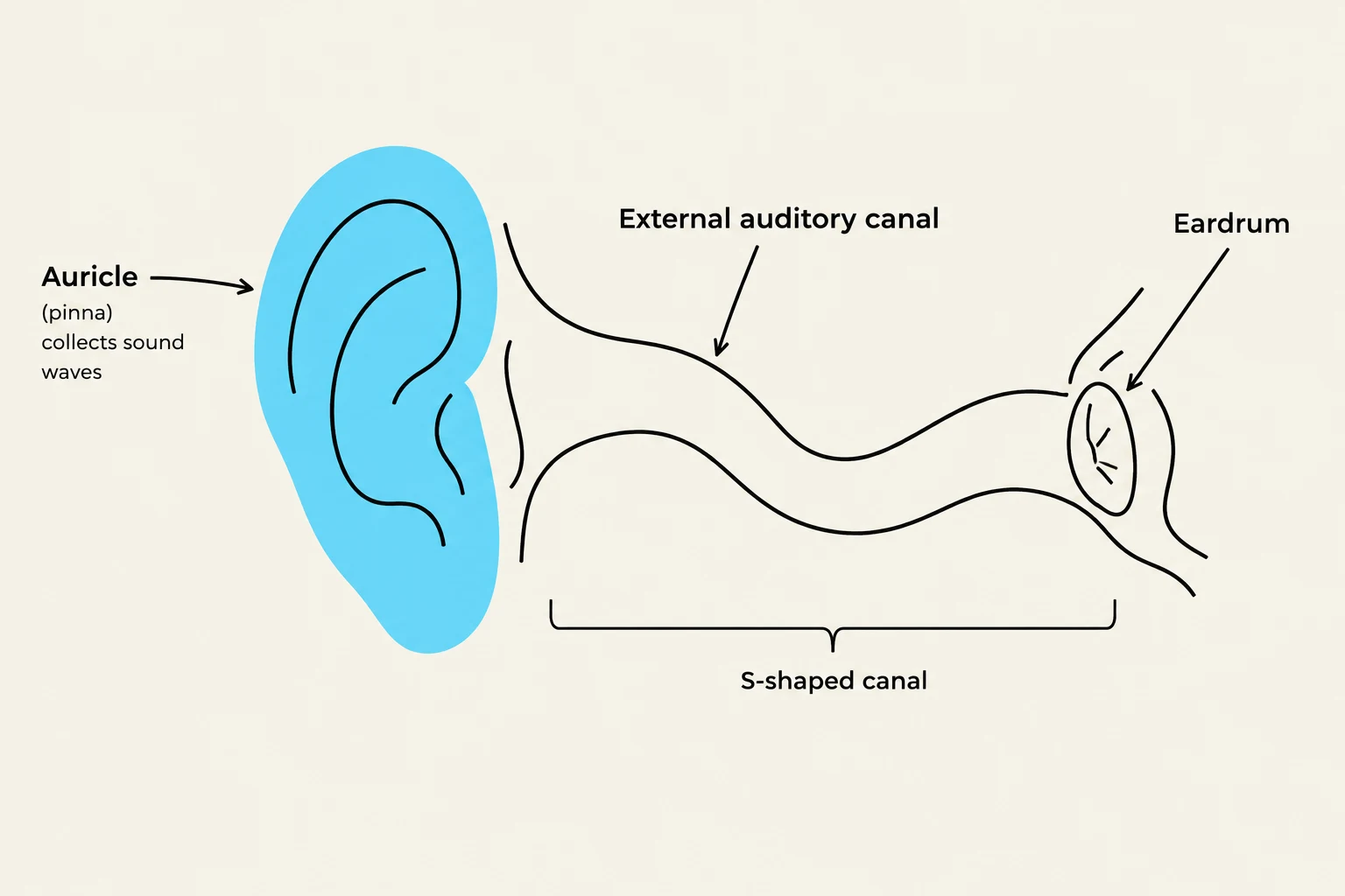
The External Ear
The external ear consists of the auricle (pinna) and the external auditory canal. The auricle is composed of elastic cartilage covered by skin and is visible without the use of instruments. The auditory canal in adults typically follows an S-shaped path. Within this canal, modified sweat glands secrete cerumen (earwax), which serves to protect the ear.
The Middle Ear
The middle ear is a small, air-filled chamber located within the temporal bone. It is separated from the external ear by the tympanic membrane (eardrum). This chamber contains the three smallest bones in the human body, known as the auditory ossicles: the malleus, incus, and stapes. A critical component of this section is the Eustachian tube, which functions to equalize air pressure on both sides of the tympanic membrane.
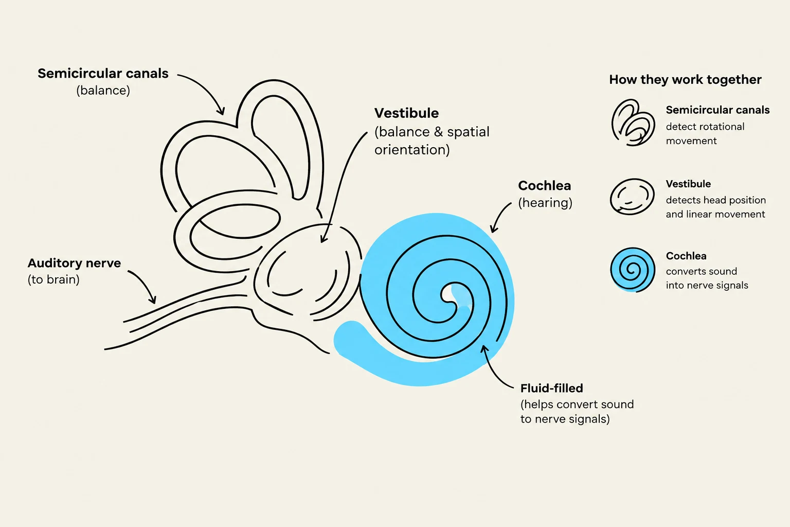
The Inner Ear
The inner ear is a fluid-filled labyrinth consisting of both a bony labyrinth and a membranous labyrinth. The bony labyrinth includes the cochlea, the vestibule, and the semicircular canals. Inside the cochlear duct lies the spiral organ of Corti, which serves as the actual sensory organ for hearing. When sound waves stimulate the hair cells within this organ, nerve impulses are sent to the brain via the acoustic nerve, which is part of cranial nerve VIII.
Keep reading for free.
Create an account to finish this reading and make it part of your study routine.
Continue reading freeAlready have an account? Log in
Updated