The human axial skeleton provides the central structural framework for the body. Among its most critical components are the vertebral column and the thoracic cage. Together, these structures serve a dual purpose: they provide the necessary rigidity to support the weight of the trunk and protect the delicate neural and visceral organs housed within. Understanding their complex anatomy is essential for grasping how the body manages movement, weight distribution, and protection.
The Vertebral Column: Structure and Function
The vertebral column is a flexible, curved structure composed of 26 irregular bones known as vertebrae. Its primary roles are to transmit the weight of the upper body to the lower limbs and to surround and protect the spinal cord.
General Vertebral Anatomy
While vertebrae vary by region, they share a common structural blueprint:
Body (Centrum): The thick, anterior portion of the bone designed for weight-bearing.
Vertebral Arch: Formed by the pedicles and laminae, this arch encloses the vertebral foramen.
Vertebral Canal: The continuous passage formed by the alignment of the vertebral foramina, which houses the spinal cord.
Intervertebral Foramina: Lateral openings located between adjacent vertebrae that allow for the exit of spinal nerves.
Anatomical illustration of a typical vertebra, highlighting its key structures such as the body, vertebral arch, spinous process, transverse process, and articular facets.
Intervertebral Discs
To prevent bone-on-bone contact and absorb shock, the vertebrae are separated by intervertebral discs. These cushionlike pads consist of two distinct parts:
Nucleus Pulposus: The inner, gelatinous core that provides elasticity and compressibility.
Anulus Fibrosus: The outer collar composed of collagen and fibrocartilage that contains the nucleus.
Clinical Note: Disc Herniation
As we age, intervertebral discs become less hydrated and less elastic. This increases the risk of a herniated disc, where the nucleus pulposus protrudes through the anulus fibrosus, potentially compressing a spinal nerve root.
Regional Vertebrae
The vertebral column is divided into five distinct regions, each specialized for different mechanical tasks:
1. Cervical Vertebrae (C1–C7)
These are the smallest and lightest vertebrae. They are characterized by transverse foramina (holes in the transverse processes) and often feature bifid spinous processes.
The Atlas (C1): A unique vertebra that lacks a body and a spinous process. It articulates with the occipital condyles of the skull, facilitating the "yes" movement of the head.
The Axis (C2): Features a prominent projection called the dens, which acts as a pivot point for the atlas, allowing for the "no" rotation of the head.
2. Thoracic Vertebrae (12 vertebrae)
These vertebrae are heart-shaped and larger than cervical vertebrae. Their defining feature is the presence of costal facets (or demifacets) on the bodies and transverse processes, which serve as articulation points for the ribs. Their spinous processes generally point inferiorly.
3. Lumbar Vertebrae (5 vertebrae)
The lumbar vertebrae are massive and kidney-shaped. Because they are located in the lower back, they receive the most significant mechanical stress from weight-bearing. Their facet orientation is specialized to allow flexion and extension while preventing excessive rotation.
4. Sacrum and Coccyx
The sacrum consists of five fused vertebrae that form the posterior wall of the pelvis. The coccyx, or tailbone, consists of 3–5 fused vertebrae at the very inferior terminus of the column.
Spinal Curvatures and Abnormalities
The adult spine is not straight; it possesses natural curvatures that increase resilience and flexibility.
Primary Curvatures: The thoracic and sacral curvatures are present at birth and give the infant spine a C-shape.
Secondary Curvatures: The cervical and lumbar curvatures develop during childhood as the individual learns to lift their head and walk.
When these curvatures deviate from the norm, several clinical conditions can arise:
Scoliosis: An abnormal lateral (side-to-side) curvature of the spine.
Kyphosis: An exaggerated posterior curvature of the thoracic spine, often resulting in a "hunchback" appearance.
Lordosis: An exaggerated anterior curvature of the lumbar spine, often called "swayback".
The Thoracic Cage
The thoracic cage is a bony and cartilaginous enclosure that protects vital organs (like the heart and lungs), supports the shoulder girdles, and provides attachment sites for various muscles. It is composed of the thoracic vertebrae posteriorly, the ribs laterally, and the sternum and costal cartilages anteriorly.
An illustration of the human rib cage, labeling the sternum, true ribs, false ribs, floating ribs, and associated anatomical landmarks.
The Sternum
The sternum is located anteriorly and features several key landmarks:
Jugular Notch: A central indentation on the superior border of the manubrium.
Sternal Angle: A horizontal ridge where the manubrium meets the body of the sternum.
Xiphisternal Joint: The point where the sternal body fuses with the xiphoid process.
Rib Structure and Classification
There are 12 pairs of ribs that all attach posteriorly to the thoracic vertebrae. They are classified based on their anterior attachment to the sternum:
True (Vertebrosternal) Ribs (1–7): These attach directly to the sternum via their own individual costal cartilages.
False (Vertebrochondral) Ribs (8–10): These attach indirectly to the sternum by joining the costal cartilage of the rib immediately above them.
Floating (Vertebral) Ribs (11–12): These have no anterior attachment to the sternum at all.
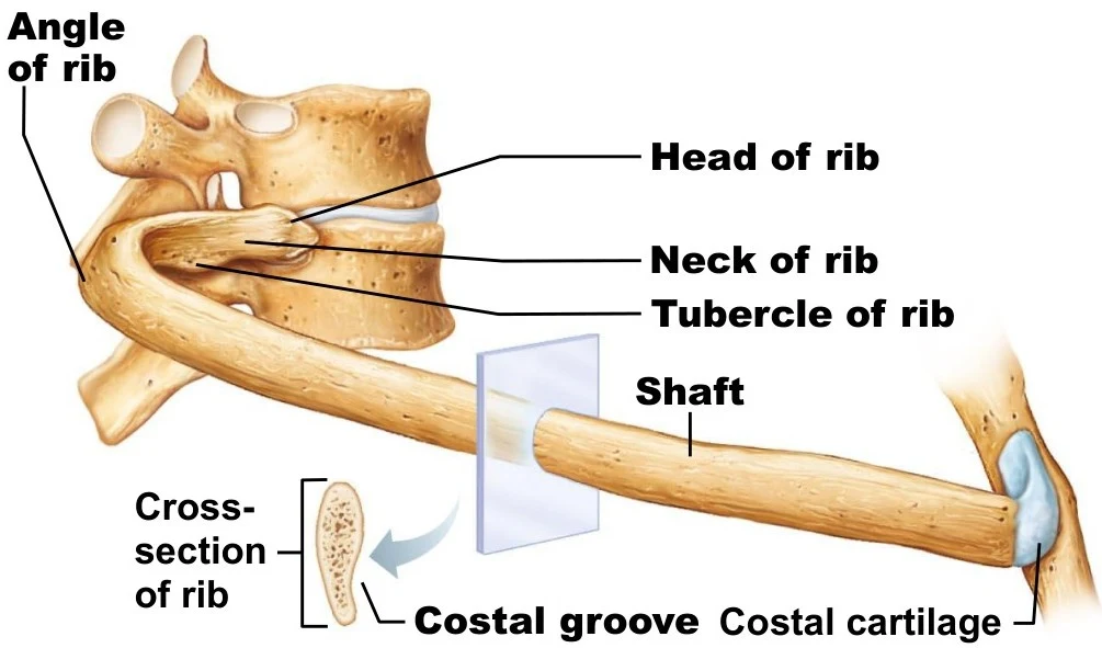
An illustration shows the anatomy of a rib articulating with a vertebra, labeling key parts such as the head, neck, tubercle, and shaft of the rib, as well as the costal groove and costal cartilage.
Exam Tip: Rib Identification
To quickly distinguish between rib types, look at the anterior connection. If it hits the sternum directly, it's True. If it "piggybacks" on another cartilage, it's False. If it's just hanging there with no front connection, it's Floating.
Keep reading for free.
Create an account to finish this reading and make it part of your study routine.